Image of figeater beetle
Description:
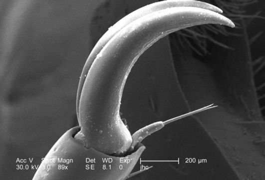
Still at a low magnification of 89X, which is twice as high as PHIL 9943, this scanning electron micrograph (SEM) again depicted some of the morphologic exoskeletal features located at the distal end of an adult figeater beetles, Cotinis mutabilis leg. Depicted here was the claw-like tip of the insects 5th tarsomere, as well as its accompanying empodium. This appendicular configuration affords the beetle a secure grasp of objects within its environmental domain, such as foliage or food. See PHIL 9943, 9945, 9947, 9948, and 9949 for additional views of these exoskeletal features.
Created: 2007
Included On The Following Pages:
- Life (creatures)
- Cellular (cellular organisms)
- Eukaryota (eukaryotes)
- Opisthokonta (opisthokonts)
- Metazoa (Animal)
- Bilateria
- Protostomia (protostomes)
- Ecdysozoa (ecdysozoans)
- Arthropoda (arthropods)
- Pancrustacea
- Hexapoda (hexapods)
- Insecta (insects)
- Pterygota (winged insects)
- Neoptera (neopteran)
- Endopterygota (endopterygotes)
- Coleoptera (beetles)
- Polyphaga
- Scarabaeiformia
- Scarabaeoidea (scarab beetle)
- Cotinis
- Cotinis mutabilis (figeater beetle)
- Scarabaeidae (scarab beetles)
- Panarthropoda
This image is not featured in any collections.
Source Information
- license
- cc-publicdomain
- photographer
- Janice Carr
- provider
- Public Health Image Library
- original
- original media file
- visit source
- partner site
- Public Health Image Library
- ID