Image of figeater beetle
Description:
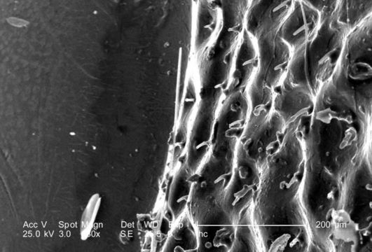
At a magnification of 130X, this scanning electron micrograph (SEM) focused on the head region of an adult figeater beetle, Cotinis mutabilis. In this particular view, the transitional area between one of the insects two compound eyes, i.e., right eye, and the vertex of its head is visualized. For even greater magnifications of the surface of the eye, see PHIL 9950, 9951, and 9952.
Created: 2007
Included On The Following Pages:
- Life (creatures)
- Cellular (cellular organisms)
- Eukaryota (eukaryotes)
- Opisthokonta (opisthokonts)
- Metazoa (Animal)
- Bilateria
- Protostomia (protostomes)
- Ecdysozoa (ecdysozoans)
- Arthropoda (arthropods)
- Pancrustacea
- Hexapoda (hexapods)
- Insecta (insects)
- Pterygota (winged insects)
- Neoptera (neopteran)
- Endopterygota (endopterygotes)
- Coleoptera (beetles)
- Polyphaga
- Scarabaeiformia
- Scarabaeoidea (scarab beetle)
- Cotinis
- Cotinis mutabilis (figeater beetle)
- Scarabaeidae (scarab beetles)
- Panarthropoda
This image is not featured in any collections.
Source Information
- license
- cc-publicdomain
- photographer
- Janice Carr
- provider
- Public Health Image Library
- original
- original media file
- visit source
- partner site
- Public Health Image Library
- ID