Image of Coccidioides immitis G. W. Stiles 1896
Description:
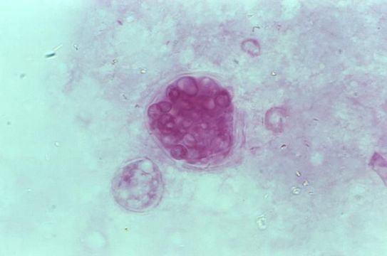
This photomicrograph revealed some of the histopathologic characteristics found within a pus specimen, which was prepared using periodic acid-Schiff (PAS), and which had been harvested from a skin lesion in a case of cutaneous coccidioidomycosis. In this particular specimen youll note the chlamydospore, or mature spherule of a Coccidioides immitis fungal organism. As the reproductive structure of this, as well as other types of fungi, this spherule is also known as a chlamydoconidium, and contains the organisms endospores.
Created: 1975
Included On The Following Pages:
- Life (creatures)
- Cellular (cellular organisms)
- Eukaryota (eukaryotes)
- Opisthokonta (opisthokonts)
- Nucletmycea
- Fungi (mushrooms, lichens, molds, yeasts and relatives)
- Dikarya
- Ascomycota (sac fungi)
- Eurotiomycetes
- Onygenales
- Onygenaceae
- Coccidioides
- Coccidioides immitis
- Pezizomycotina
- Eurotiomycetidae
This image is not featured in any collections.
Source Information
- license
- cc-publicdomain
- provider
- Public Health Image Library
- original
- original media file
- visit source
- partner site
- Public Health Image Library
- ID