Image of Brown Recluse
Description:
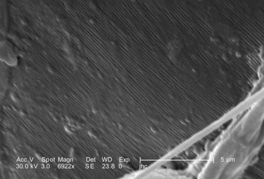
Under a high magnification of 6922x this scanning electron micrograph (SEM) depicted the striated texture found on the exoskeletal surface of a venomous brown recluse spider, Loxosceles reclusa, found inhabiting a Kentucky farm. As arthropods, spiders possess an exoskeleton composed of chitin, which is a molecule made up of bound units of acetylglucosamine, joined in such a way as to allow for increased points at which hydrogen bonding can occur. In this way chitin provides increased strength, and durability as an exoskeletal foundation. L. reclusa is sometimes referred to as the violin or fiddle spider, for on its cephalothorax one will see what appears to be coloration in the shape of these stringed instruments, which is quite evident in the color photograph PHIL 1125, depicting a live specimen. Also see PHIL 2224, and 6268 for additional brown recluse images.
Created: 2007
Included On The Following Pages:
- Life (creatures)
- Cellular (cellular organisms)
- Eukaryota (eukaryotes)
- Opisthokonta (opisthokonts)
- Metazoa (Animal)
- Bilateria
- Protostomia (protostomes)
- Ecdysozoa (ecdysozoans)
- Arthropoda (arthropods)
- Chelicerata (chelicerates)
- Arachnida (arachnids)
- Araneae (spiders)
- Opisthothelae
- Araneomorphae
- Haplogynae
- Sicariidae (six-eyed brown spiders)
- Loxosceles (Recluse Spiders)
- Loxosceles reclusa (Brown Recluse)
- Panarthropoda
This image is not featured in any collections.
Source Information
- license
- cc-publicdomain
- photographer
- Janice Haney Carr
- provider
- Public Health Image Library
- original
- original media file
- visit source
- partner site
- Public Health Image Library
- ID