Image of Dasymutilla Ashmead 1899
Description:
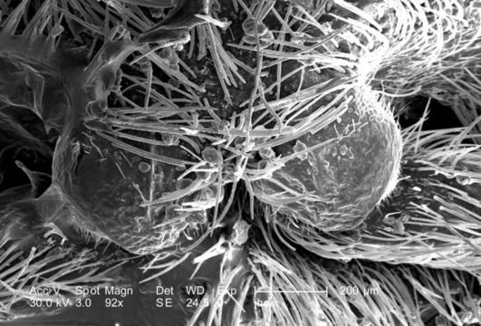
Double the magnification of PHIL 9898, at 92X, this scanning electron micrograph (SEM) showed the head region from an anterior view of a female velvet ant, Dasymutilla sp.. Note the two anteriorly-placed antennae with their rounded "scapes" that are the most apparent head appendages. Like the antennae, the numerous hairs or setae adorning almost all of the insects exterior surfaces, act as sensory structures, supplying the organism with information about its environmental parameters. The jointed legs, from which the insects Phylum Arthropoda is derived, i.e., Arthro = jointed, and poda leg, are also partially visible, emanating from the thoracic region.
Created: 2007
Included On The Following Pages:
- Life (creatures)
- Cellular (cellular organisms)
- Eukaryota (eukaryotes)
- Opisthokonta (opisthokonts)
- Metazoa (Animal)
- Bilateria
- Protostomia (protostomes)
- Ecdysozoa (ecdysozoans)
- Arthropoda (arthropods)
- Pancrustacea
- Hexapoda (hexapods)
- Insecta (insects)
- Pterygota (winged insects)
- Neoptera (neopteran)
- Endopterygota (endopterygotes)
- Hymenoptera (wasps, bees, and ants)
- Apocrita (wasp)
- Aculeata
- Vespoidea (Yellowjackets and Hornets, Paper Wasps; Potter, Mason and Pollen Wasps and allies)
- Mutillidae (velvet ants)
- Dasymutilla
- Panarthropoda
This image is not featured in any collections.
Source Information
- license
- cc-publicdomain
- photographer
- Janice Carr
- provider
- Public Health Image Library
- original
- original media file
- visit source
- partner site
- Public Health Image Library
- ID