Image of Niphargus plurispinosus Hudec & Mock 2014
Description:
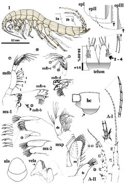
Figure 2.Niphargus plurispinosus sp. n.: 1 male, general view; 1a-1b dorso-later thorns; mdb - mandibula and details of mdb-a left incisor and lacina mobilis; mdb-b) two setae between bisserated thorns; mdb-c setae pattern on distal segment of mdb-palp; mdb-d, right incisor and lacina mobilis; mx-1 1st maxilla; mx-2 2nd maxilla; ula upper lip; vela ventral labium; mxp maxilliped: in inner segment os outer segment; ds distal segment of palp; epI-epIII epimeral plate I-III; A-I 1st antenna; A-II antenna; hc head capsula, left lateral view; telson, dorsal view. Not scaled, except of the general view of the male.
Included On The Following Pages:
- Life
- Cellular
- Eukaryota (eukaryotes)
- Opisthokonta (opisthokonts)
- Metazoa (animals)
- Bilateria
- Protostomia (protostomes)
- Ecdysozoa (ecdysozoans)
- Arthropoda (arthropods)
- Pancrustacea
- Multicrustacea (typical crustaceans)
- Malacostraca (malacostracans)
- Eumalacostraca
- Peracarida (peracarids)
- Amphipoda (amphipods)
- Senticaudata
- Gammarida
- Crangonyctidira
- Crangonyctoidea
- Niphargidae
- Niphargus
- Niphargus plurispinosus
- Panarthropoda
This image is not featured in any collections.
Source Information
- license
- cc-by-3.0
- copyright
- Igor Hudec, Andrej Mock
- provider
- Subterranean Biology
- original
- original media file
- visit source
- partner site
- Subterranean Biology (archived)
- ID