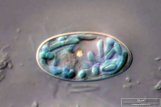
Image of Glaucocystales
Description:
Scale bars indicate 10 µm.
Four images.
Pairs of images of individual cells. The first shows a synoptic view of the anterior cell wall, the second is an optical cross-section at the level of the cell nucleus.
Please click on < or > on the image edges or on the dots at the bottom edge of the images to browse through the slides!
Place name: Protist culture SAG Göttingen (Germany)
Latitude: 51.53138716 Longitude: 9.939193725
Microscope Zeiss Axioplan, camera Canon EOLS 600D. DOF images.
Sample material courtesy of AG Boenigk, University Duisburg-Essen.
© Wolfgang Bettighofer,
images under Creative Commons License V 3.0 (CC BY-NC-SA).
For permission to use of (high resolution) images please contact postmaster@protisten.de.
For further information about the image, please click here: Link to protisten.de page
Included On The Following Pages:
- Life (biota)
- Cellular
- Eukaryota (eukaryotes)
- Archaeplastida (plants)
- Glaucophyta (glaucophytes)
- Glaucocystophyceae
- Glaucocystales
- Glaucocystaceae
- Glaucocystis
- Glaucocystis nostochinearum
This image is not featured in any collections.
Source Information
- license
- cc-by-nc-sa-3.0
- copyright
- Wolfgang Bettighofer
- creator
- Wolfgang Bettighofer [email]
- original
- original media file
- visit source
- partner site
- protisten.de
- ID