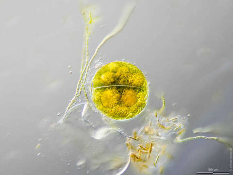
Image of Eremosphaera viridis
Description:
Sampling date 10/2019. Scale bars indicate 250 µm (1), 100 µm (2, 3).
Three images. Advanced stages of development of vegetative cells from cystozygotes.
Please click on < or > on the image edges or on the dots at the bottom edge of the images to browse through the slides!
Place name: Bog Waasenmoos Pass Thurn near Mittersil (Tyrol, Austria)
Latitude: 47.30234117 Longitude: 12.41751194
Stereo microscope Olympus SZX16, camera Olympus OM-D M5 MKII. DOF images.
© Wolfgang Bettighofer,
images under Creative Commons License V 3.0 (CC BY-NC-SA).
For permission to use of (high resolution) images please contact postmaster@protisten.de.
For further information about the image, please click here: Link to protisten.de page
Included On The Following Pages:
- Life
- Cellular
- Eukaryota (eukaryotes)
- Archaeplastida (plants)
- Chloroplastida
- Chlorophyta (chlorophytes)
- Trebouxiophyceae
- Chlorellales
- Oocystaceae
- Eremosphaeroideae
- Eremosphaera
- Eremosphaera viridis
This image is not featured in any collections.
Source Information
- license
- cc-by-nc-sa-3.0
- copyright
- Wolfgang Bettighofer
- creator
- Wolfgang Bettighofer [email]
- original
- original media file
- visit source
- partner site
- protisten.de
- ID