Image of Ceramium diaphanum
Description:
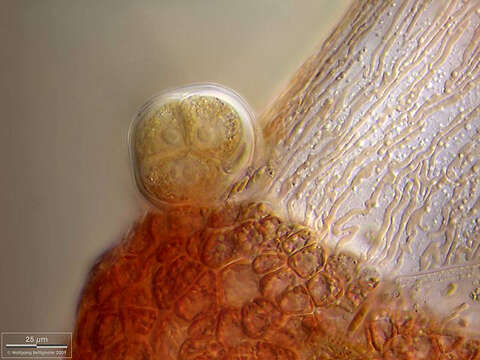
Scale bars indicate 200 µm (1), 100 µm (2), 50 µm (3, 4), 25 µm (5).
Five images.
First:A dichotomously branching red alga of the genus Ceramium. The image shows axial cells that are partially surrounded by cortical cells (red belts). Filament hairs emerge from the cortical cells.Second:Close-up.Third:The cortical cells have round chloroplasts, while the chloroplasts of the axial cells are worm-shaped.Fourth:First cells of an adventitious shoot of Ceramium diaphanum , covered by a thick gelatinous layer (agar-like pectin).Fifth:This detailed view of a thallus segment shows cells of the two types found: a long axial cell (light, with worm-shaped rhodoplasts) and many small, round, reddish, cortical cells with their lens-shaped rhodoplasts. In the rounded outgrowth (tetrasporangium) three of the four cells with their nuclei are visible.
Please click on < or > on the image edges or on the dots at the bottom edge of the images to browse through the slides!
Place name: Hiddensee Bodden (Germany)
Latitude: 54.582633 Longitude: 13.115051
Microscope Zeiss Universal, camera Olympus C7070WZ. DOF images.
© Wolfgang Bettighofer,
images under Creative Commons License V 3.0 (CC BY-NC-SA).
For permission to use of (high resolution) images please contact postmaster@protisten.de.
For further information about the image, please click here: Link to protisten.de page
Included On The Following Pages:
- Life
- Cellular
- Eukaryota (eukaryotes)
- Archaeplastida (plants)
- Rhodophyta (red algae)
- Florideophyceae (Florideae)
- Rhodymeniophycidae
- Ceramiales
- Ceramiaceae
- Ceramium
- Ceramium diaphanum
This image is not featured in any collections.
Source Information
- license
- cc-by-nc-sa-3.0
- copyright
- Wolfgang Bettighofer
- creator
- Wolfgang Bettighofer [email]
- original
- original media file
- visit source
- partner site
- protisten.de
- ID