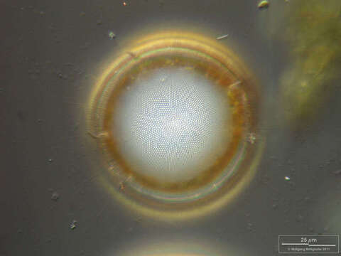
Image of Thalassiosira lundiana
Description:
Sampling date 04/2011. Scale bars indicate 25 µm.
Two images.
First:Silicious processes (the labiate and the occluded ones) are visible.Second:Labiate processes and chitinous spines (typical for the Thalassiosirales) are visible.Please click on < or > on the image edges or on the dots at the bottom edge of the images to browse through the slides!
Place name: North Sea around Heligoland
Latitude: 54.186311 Longitude: 7.895034
Microscope Zeiss Universal, camera Olympus C7070WZ. DOF images.
© Wolfgang Bettighofer,
images under Creative Commons License V 3.0 (CC BY-NC-SA).
For permission to use of (high resolution) images please contact postmaster@protisten.de.
For further information about the image, please click here: Link to protisten.de page
Included On The Following Pages:
- Life
- Cellular
- Eukaryota (eukaryotes)
- SAR (Stramenopiles, Alveolates, Rhizaria)
- Stramenopiles (heterokont)
- Ochrophyta (Ochrophyte)
- Bacillariophyta (diatoms)
- Coscinodiscophyceae
- Thalassiosirophycidae
- Thalassiosirales
- Thalassiosiraceae
- Thalassiosira
- Thalassiosira lundiana
This image is not featured in any collections.
Source Information
- license
- cc-by-nc-sa-3.0
- copyright
- Wolfgang Bettighofer
- creator
- Wolfgang Bettighofer [email]
- original
- original media file
- visit source
- partner site
- protisten.de
- ID