Image of Zoothamnium arbuscula
Description:
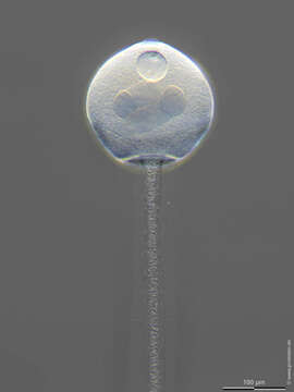
Scale bars indicate 100 µm.
Specimen grown in a Petri dish on the edge of a microscope slide.
An image couple, the first without and the second with marking arrows.
The images show a initial cell for a Zoothamnium tree. In this ciliate species, only special cells, the macrozooids, can separate themselves from the mother tree as swarmers and form new trees.
Macronucleus (arrow), contractile vacuole (arrowhead), contractile stalk (double arrowhead).
Please click on < or > on the image edges or on the dots at the bottom edge of the images to browse through the slides!
Place name: Creek in Oder valley 100 km north east of Berlin (Germany)
Latitude: 53.135032 Longitude: 14.348738
Stereo microscope Olympus SZX16/Planapo 2.0x, camera Olympus OM-D M5 MKII. DOF images.
© Wolfgang Bettighofer,
images under Creative Commons License V 3.0 (CC BY-NC-SA).
For permission to use of (high resolution) images please contact postmaster@protisten.de.
For further information about the image, please click here: Link to protisten.de page
Included On The Following Pages:
- Life
- Cellular
- Eukaryota (eukaryotes)
- SAR (Stramenopiles, Alveolates, Rhizaria)
- Alveolata (alveolates)
- Ciliophora (ciliates)
- Intramacronucleata
- Oligohymenophorea
- Peritrichia
- Sessilida
- Zoothamniidae
- Zoothamnium
- Zoothamnium arbuscula
This image is not featured in any collections.
Source Information
- license
- cc-by-nc-sa-3.0
- copyright
- Wolfgang Bettighofer
- creator
- Wolfgang Bettighofer [email]
- original
- original media file
- visit source
- partner site
- protisten.de
- ID