Infraciliature
Description:
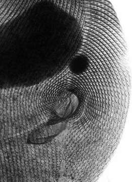
Detail of the oral apparatus and surrounding infraciliature (anterior ventral aspect) of the hymenostome ciliate Paramecium caudatum (Ehrenberg, 1833). The preoral and postoral sutures (lines of convergence of kinetal fields) can be clearly seen. The kinetids with their kinetodesmal fibers are clearly visible. The kinetodesmal fibers are periodically striated bundles of fibrils arising near the base of somatic kinetids (the posterior one if kinetids are paired) in ciliates. The kinetodesmal fibers extend anteriorly and to the right of their kinety (this is the Law of desmodexy). This provides a means of determining an anterior, posterior and right/left orientation in ciliates. Here the kinetodesmal fibers are longer than the interkinetal distance and therefore overlap giving the appearance of a longitudinal interkinetal line. The macronucleus and the single large micronucleus are seen anterior to the oral aperture. Silver carbonate stain (see Foissner, W.Europ. J. Protistol.27,313-330;1991). Specimen collected from freshwater pond near Boise, Idaho July 2004. Brightfield.
Included On The Following Pages:
- Life
- Cellular
- Eukaryota (eukaryotes)
- SAR (Stramenopiles, Alveolates, Rhizaria)
- Alveolata (alveolates)
- Ciliophora (ciliates)
- Intramacronucleata
- Oligohymenophorea
- Peniculida (Peniculid)
- Parameciidae
- Paramecium (slipper animalcules)
- Paramecium caudatum (slipper animalcule)
This image is not featured in any collections.
Source Information
- license
- cc-by-nc
- author
- William Bourland
- provider
- micro*scope
- original
- original media file
- visit source
- partner site
- micro*scope
- ID