Infraciliature
Description:
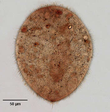
Infraciliature (ventral) of the ciliate, Ophryoglena utriculariae (Kahl, 1831). The numerous closely spaced longitudinal kineties on the right and left of the oral aperture converge at a sigmoid preoral suture. At least 15 postoral kineties terminate at the oral aperture. There is a single long caudal cilium. The dense inner ciliary rows of the buccal opening are seen here as a brown crescent in the concavity of which the refractile "watchglass organelle" (Lieberkühn's organelle) is located (seen well here).There is one right lateral contractile vacuole (not seen here). The large ellipsoid macronucleus is seen here with its anterior end at the posterior margin of the oral aperture. Collected from a freshwater pond near Boise, Idaho. Silver carbonate method (see Foissner, W.Europ. J. Protistol.27,313-330;1991). Brightfield.
Included On The Following Pages:
- Life
- Cellular
- Eukaryota (eukaryotes)
- SAR (Stramenopiles, Alveolates, Rhizaria)
- Alveolata (alveolates)
- Ciliophora (ciliates)
- Intramacronucleata
- Oligohymenophorea
- Hymenostomatida
- Ophryoglenina
- Ophryoglena
- Ophryoglena utriculariae
This image is not featured in any collections.
Source Information
- license
- cc-by-nc
- author
- William Bourland
- provider
- micro*scope
- original
- original media file
- visit source
- partner site
- micro*scope
- ID