portrait
Description:
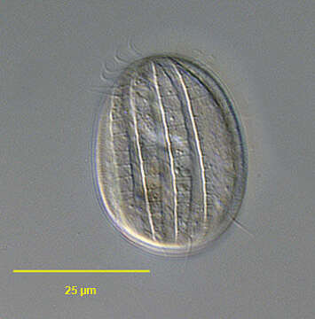
Portrait (dorsolateral view) of the microthoracid ciliate Pseudomicrothorax agilis (Mermod,1914).The cell outline is oval. Laterally compressed. The right and left sides of the inflexible pellicle have curved longitudinal ciliated grooves separated by broader flat ridges. The ridges are transversely striated with extrusomes between the striations.Somatic ciliature restricted to 12 longitudinal kineties more on the right than the left surface. The oral aperture is in anterior 1/3 of body in a shallow depression. The cytopharynx is supported by fine transverse trichites. There are 3 short membranelles on the left of the cytostome and a short undulating membrane on its right. There is a small group of unciliated basal bodies posterior to the undulating membrane. This is a primordial stomatogenic field. There are many fusiform peripheral extrusomes. Macronucleus elongate ovoid, centrally placed with single micronucleus. The contractile vacuole is centrally located. P. agilis is smaller with fewer longitudinal kineties than the similar P. dubius. Collected from a freshwater stream near Boise, Idaho. DIC.
Included On The Following Pages:
- Life (biota)
- Cellular
- Eukaryota (eukaryotes)
- SAR (Stramenopiles, Alveolates, Rhizaria)
- Alveolata (alveolates)
- Ciliophora (ciliates)
- Intramacronucleata
- Nassophorea
- Microthoracida
- Pseudomicrothoracidae
- Pseudomicrothorax
- Pseudomicrothorax agilis
This image is not featured in any collections.
Source Information
- license
- cc-by-nc
- author
- William Bourland
- provider
- micro*scope
- original
- original media file
- visit source
- partner site
- micro*scope
- ID