Ventral infraciliature
Description:
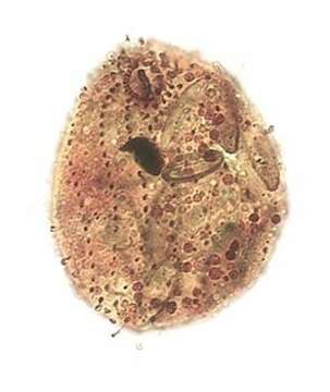
Ventral infraciliature of the colpodid ciliate, Platyophrya vorax (Kahl,1926). The flask-shaped cells are very flexible. The relatively small ovoid anterior cytostome is subapical. There is a right paraoral membrane composed of dikinetids and four rectangular left adoral membranelles (seen here). There is a postoral "pseudomembrane" consisting of closely spaced anterior dikinetids of somatic kineties (not well preserved in this specimen). The dark vertical band in the center of the cytostome probably represents densely staining pharyngeal fibers seen end-on.The right side is more densely ciliated than the left. The slightly spiralled somatic kineties lie in shallow cortical furrows. The central spherical macronucleus has a small central nucleolus and an adjacent micronucleus. The single contractile vacuole is subterminal posteriorly. Ingested diatoms are visible here.P. vorax lacks endosymbiotic algae (present in P. sphagni which also has more numerous adoral membranelles and somatic kineties).Collected from temporary puddles with heavy growth of diatoms in a meadow near Boise, Idaho. 43°41'45.09"N 116°13'55.29"W elev.3191 ft. March 2006. Stained by the silver carbonate technique (see Foissner, W. Europ. J. Protistol., 27:313-330;1991).Brightfield.
Included On The Following Pages:
- Life (creatures)
- Cellular (cellular organisms)
- Eukaryota (eukaryotes)
- SAR (Stramenopiles, Alveolates, Rhizaria)
- Alveolata (alveolates)
- Ciliophora (ciliates)
- Intramacronucleata
- Colpodea
- Cyrtolophosidida
- Platyophryidae
- Platyophrya
- Platyophrya vorax
This image is not featured in any collections.
Source Information
- license
- cc-by-nc
- author
- William Bourland
- provider
- micro*scope
- original
- original media file
- visit source
- partner site
- micro*scope
- ID