Infraciliature in division
Description:
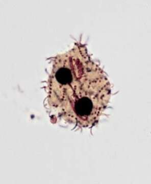
Ventral infraciliature of Cyrtolophosis mucicola (Stokes,1885)in middle division. The paraoral membrane is seen to the viewer's right and the adoral membranelles to the left.A very short "oblique kinety" is present between the anterior end of the paraoral membrane and the most anterior adoral membranelle. The sparse (~10) slightly spiralled somatic kineties are visible.The oral apparatus of the forming posterior daughter cell (opisthe) is obscured by its densley stained macronucleus. Collected from a freshwater pond near Boise, Idaho.June 2005). Stained by the silver carbonate technique (see Foissner, W. Europ. J. Protistol., 27:313-330;1991). Brightfield.
Included On The Following Pages:
- Life (creatures)
- Cellular (cellular organisms)
- Eukaryota (eukaryotes)
- SAR (Stramenopiles, Alveolates, Rhizaria)
- Alveolata (alveolates)
- Ciliophora (ciliates)
- Intramacronucleata
- Colpodea
- Cyrtolophosidida
- Cyrtolophosididae
- Cyrtolophosis
- Cyrtolophosis mucicola
This image is not featured in any collections.
Source Information
- license
- cc-by-nc
- author
- William Bourland
- provider
- micro*scope
- original
- original media file
- visit source
- partner site
- micro*scope
- ID