Pseudo-nitzschia plurisecta
Description:
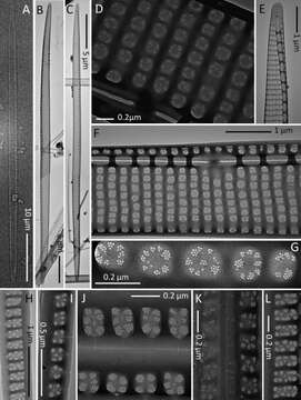
(A) Light micrographs. (B–L) TEM micrographs. (A) Whole valve. (B and C) Complete valve of different shapes. (D) Detail of the central nodule and striae with a row of poroids split in several sectors. (E) Apical part of a valve. (F) Central part of a valve showing the central nodule, the poroid arrangement, and the proximal and distal mantles. (G) Detail of the poroid structure. (H and L) Detail of the valvocopula. (I–K) Different cingular bands. (A–C, F–H, J, and K) Strain Ner-F1. (D) Strain Ner-J6. (E, I and L) Strain Ner-G4.
Included On The Following Pages:
- Life
- Cellular
- Eukaryota (eukaryotes)
- SAR (Stramenopiles, Alveolates, Rhizaria)
- Stramenopiles (heterokont)
- Ochrophyta (Ochrophyte)
- Bacillariophyta (diatoms)
- Bacillariophyceae
- Bacillariophycidae
- Bacillariales
- Bacillariaceae
- Pseudo-nitzschia
- Pseudo-nitzschia plurisecta
This image is not featured in any collections.
Source Information
- license
- cc-by-nc-sa-4.0
- copyright
- WoRMS Editorial Board
- contributor
- Orive, Emma
- original
- original media file
- visit source
- partner site
- World Register of Marine Species
- ID