1901 Die Entwicklungsgeschichte der Scolopender t2 f08-13
Description:
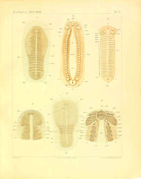
Summary.mw-parser-output table.commons-file-information-table,.mw-parser-output.fileinfotpl-type-information{border:1px solid #a2a9b1;background-color:#f8f9fa;padding:5px;font-size:95%;border-spacing:2px;box-sizing:border-box;margin:0;width:100%}.mw-parser-output table.commons-file-information-table>tbody>tr,.mw-parser-output.fileinfotpl-type-information>tbody>tr{vertical-align:top}.mw-parser-output table.commons-file-information-table>tbody>tr>td,.mw-parser-output table.commons-file-information-table>tbody>tr>th,.mw-parser-output.fileinfotpl-type-information>tbody>tr>td,.mw-parser-output.fileinfotpl-type-information>tbody>tr>th{padding:4px}.mw-parser-output.fileinfo-paramfield{background:#ccf;text-align:right;padding-right:0.4em;width:15%;font-weight:bold}.mw-parser-output.commons-file-information-table+table.commons-file-information-table,.mw-parser-output.commons-file-information-table+div.commons-file-information-table>table{border-top:0;padding-top:0;margin-top:-8px}@media only screen and (max-width:719px){.mw-parser-output table.commons-file-information-table,.mw-parser-output.commons-file-information-table.fileinfotpl-type-information{border-spacing:0;padding:0;word-break:break-word;width:100%!important}.mw-parser-output.commons-file-information-table>tbody,.mw-parser-output.fileinfotpl-type-information>tbody{display:block}.mw-parser-output.commons-file-information-table>tbody>tr>td,.mw-parser-output.commons-file-information-table>tbody>tr>th,.mw-parser-output.fileinfotpl-type-information>tbody>tr>td,.mw-parser-output.fileinfotpl-type-information>tbody>tr>th{padding:0.2em 0.4em;text-align:left;text-align:start}.mw-parser-output.commons-file-information-table>tbody>tr,.mw-parser-output.fileinfotpl-type-information>tbody>tr{display:flex;flex-direction:column}.mw-parser-output.commons-file-information-table+table.commons-file-information-table,.mw-parser-output.commons-file-information-table+div.commons-file-information-table>table{margin-top:-1px}.mw-parser-output.fileinfo-paramfield{box-sizing:border-box;flex:1 0 100%;width:100%}} Description: Deutsch: Fig. 8. Junger in der Segmentierung begriffener Keimstreifen von Scolopendra cingulata. Eine An- zahl von Metameren ist bereits ziemlich deutlich abgegrenzt. Die noch etwas unregelmässigen und verschwommenen dunkleren Konturen zur Seite des hellen medianen Ventralstreifens werden durch das ungleichmässig verteilte, in der Tiefe gelegene aber durchschimmernde Mesoderm hervorgerufen. Vorn ist die Mundöffnung bereits angelegt. Vergr. 44. Fig. 9. Ausgewachsener Keimstreifen von Scolopendra cingulata während des Auseinanderweichens der lateralen Körperhälften. Vergr. 25. Fig. 10. Keimstreifen von Scolopendra cingulata nach dem Auftreten sämtlicher Rumpfsegmente. Die präantennalen Gliedmassen sind (pran) als quere, vor den Antennen gelegene wulstformige Verdickungen schon angedeutet. Vergr. ^^. Fig. 11. Kopf einer Embryonalanlage von Scolopendra cingulata. Die Antennenanlagen sind durch eine tiefe Furche von dem darauf folgenden Kopfabschnitt getrennt. In der Kieferregion ist das Mandibelsegment noch ziemlich undeutlich, während die beiden Muxillensegmente und das Segment der Maxillarfüsse bereits besser abgegrenzt sind. Vergr. 50. Fig. 1 2. Embryonalanlage von Scolopendra cingulata nach dem Auftreten der drei ersten Metameren. Der Vorderkörper ist noch nicht scharf gegen das umgebende Blastoderm abgesetzt. Das quere Band hinter der Mundöffnung wird durch das hindurchschimmernde Mesoderm veranlasst. Vergr. 46. Fig. 13. Kopf eines ausgebildeten Keimstreifens von Scolopendra cingulata. Vergr. 62. Date: 1901. Source: Heymons, Richard. (1901). Die Entwicklungsgeschichte der Scolopender. Stuttgart: E. Nägele. doi:10.5962/bhl.title.1587. Author: Richard Heymons.
Included On The Following Pages:
- Life (creatures)
- Cellular (cellular organisms)
- Eukaryota (eukaryotes)
- Opisthokonta (opisthokonts)
- Metazoa (Animal)
- Bilateria
- Protostomia (protostomes)
- Ecdysozoa (ecdysozoans)
- Arthropoda (arthropods)
- Myriapoda (myriapods)
- Chilopoda (centipedes)
- Pleurostigmophora
- Scolopendromorpha (Bark Centipedes)
- Scolopendridae
- Scolopendrinae
- Scolopendra (Centipede)
- Scolopendra cingulata (Mediterranean banded centipede)
This image is not featured in any collections.
Source Information
- license
- cc-publicdomain
- creator
- Richard Heymons
- source
- Heymons, Richard. (1901). Die Entwicklungsgeschichte der Scolopender. Stuttgart: E. Nägele. doi:10.5962/bhl.title.1587
- original
- original media file
- visit source
- partner site
- Wikimedia Commons
- ID