Detail of Sea Urchin Skeleton (31776781367)
Description:
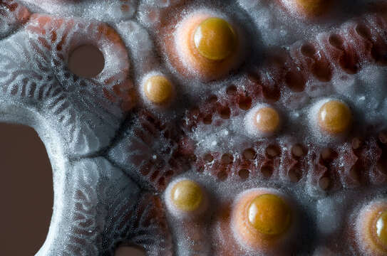
Description: The image shows a detail of the test (=shell, skeleton) of a symmetric sea urchin with a diameter of about 4 cm. The hole on the left is the anus (on the upper side of the sea urchin). It is surrounded by the apical disc containing a total of five holes (gonopores), through which eggs and sperm are released. The yellow spots are the sockets of the spines. In between are rows of holes for the tube feet protruding between the spines. Mitutoyo M-Plan Apo 5x microscope lens. The long edge of the image is actually only about 7 mm long. Focus stack with 40 steps. I used the electronically controlled focusing rack Novoflex Castel Micro and the HeliconFocus Pro software for stacking with Method A (Radius 1, Smoothing 1). Contrast slightly enhanced in Photoshop. Date: 10 January 2019, 21:54. Source: Detail of Sea Urchin Skeleton. Author: Bernd Thaller from Graz, Austria.
Included On The Following Pages:
- Life (creatures)
- Cellular (cellular organisms)
- Eukaryota (eukaryotes)
- Opisthokonta (opisthokonts)
- Metazoa (Animal)
- Bilateria
- Deuterostomia (deuterostomes)
- Echinodermata (echinoderms)
- Echinozoa
- Echinoidea (sea urchins)
This image is not featured in any collections.
Source Information
- license
- cc-by-3.0
- copyright
- Bernd Thaller|sourceurl=https://flickr.com/photos/44296132@N06/31776781367%7Carchive=%7Creviewdate=2020-08-31 09:39:57|reviewlicense=cc-by-2.0|reviewer=FlickreviewR 2
- original
- original media file
- visit source
- partner site
- Wikimedia Commons
- ID
.jpg)
.jpg)