Journal.pone.0235342.g003
Description:
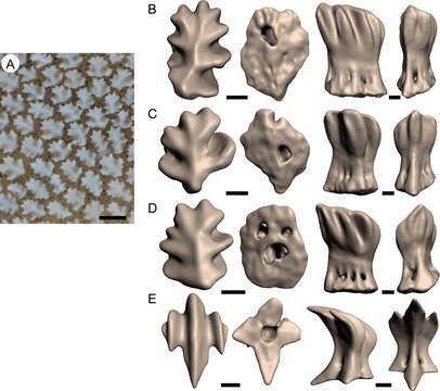
Description: English: Fig 3. Morphology of each eye denticle of the whale shark. A. Close-up of aggregated eye denticles. B–D. 3D reconstruction of eye denticles obtained from computed tomography data, showing the morphological variations. E. Dermal denticle from the skin just above of the eyeball. In B–E, apical, basal, lateral, and posterior views from left to right. Scale bars = 500 μm in A and 100 μm in B–E. See supporting information S1 Video. Date: 14 April 2020, 06:07:27. Source: https://doi.org/10.1371/journal.pone.0235342. Author: Tomita T, Murakumo K, Komoto S, Dove A, Kino M, Miyamoto K, et al. (2020) Armored eyes of the whale shark. PLoS ONE 15(6): e0235342. https://doi.org/10.1371/journal.pone.0235342.
Included On The Following Pages:
- Life (creatures)
- Cellular (cellular organisms)
- Eukaryota (eukaryotes)
- Opisthokonta (opisthokonts)
- Metazoa (Animal)
- Bilateria
- Deuterostomia (deuterostomes)
- Chordata (Chordates)
- Vertebrata (vertebrates)
- Gnathostomata (jawed fish)
- Chondrichthyes (cartilaginous fishes)
- Elasmobranchii ("sharks, skates and rays")
- Selachii (modern sharks)
- Orectolobiformes (carpet sharks)
- Rhincodontidae (whale sharks)
- Rhincodon
- Rhincodon typus (Whale Shark)
This image is not featured in any collections.
Source Information
- license
- cc-by-3.0
- copyright
- Tomita T, Murakumo K, Komoto S, Dove A, Kino M, Miyamoto K, et al. (2020) Armored eyes of the whale shark. PLoS ONE 15(6): e0235342. https://doi.org/10.1371/journal.pone.0235342
- original
- original media file
- visit source
- partner site
- Wikimedia Commons
- ID