Journal.pone.0235342.g002
Description:
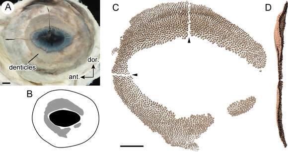
Description: English: Fig 2. Eye denticles of the whale shark. A. Distal view of left eyeball (OCF-P04248). B. Line drawing of OCF-P04248, showing the distribution of the eye denticles (gray area). C. Three-dimensional image of eye denticle aggregation obtained from computed tomography data. Horizontal and vertical lines of no denticle area (arrowheads) are artifacts. D. Posterior view of panel C. Scale bars = 0.5 cm. Date: 13 April 2020, 18:53:27. Source: https://doi.org/10.1371/journal.pone.0235342. Author: Tomita T, Murakumo K, Komoto S, Dove A, Kino M, Miyamoto K, et al. (2020) Armored eyes of the whale shark. PLoS ONE 15(6): e0235342. https://doi.org/10.1371/journal.pone.0235342.
Included On The Following Pages:
- Life (creatures)
- Cellular (cellular organisms)
- Eukaryota (eukaryotes)
- Opisthokonta (opisthokonts)
- Metazoa (Animal)
- Bilateria
- Deuterostomia (deuterostomes)
- Chordata (Chordates)
- Vertebrata (vertebrates)
- Gnathostomata (jawed fish)
- Chondrichthyes (cartilaginous fishes)
- Elasmobranchii ("sharks, skates and rays")
- Selachii (modern sharks)
- Orectolobiformes (carpet sharks)
- Rhincodontidae (whale sharks)
- Rhincodon
- Rhincodon typus (Whale Shark)
This image is not featured in any collections.
Source Information
- license
- cc-by-3.0
- copyright
- Tomita T, Murakumo K, Komoto S, Dove A, Kino M, Miyamoto K, et al. (2020) Armored eyes of the whale shark. PLoS ONE 15(6): e0235342. https://doi.org/10.1371/journal.pone.0235342
- original
- original media file
- visit source
- partner site
- Wikimedia Commons
- ID