Hhv-6- tissue culture
Description:
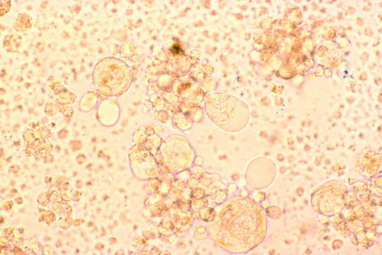
Summary.mw-parser-output table.commons-file-information-table,.mw-parser-output.fileinfotpl-type-information{border:1px solid #a2a9b1;background-color:#f8f9fa;padding:5px;font-size:95%;border-spacing:2px;box-sizing:border-box;margin:0;width:100%}.mw-parser-output table.commons-file-information-table>tbody>tr,.mw-parser-output.fileinfotpl-type-information>tbody>tr{vertical-align:top}.mw-parser-output table.commons-file-information-table>tbody>tr>td,.mw-parser-output table.commons-file-information-table>tbody>tr>th,.mw-parser-output.fileinfotpl-type-information>tbody>tr>td,.mw-parser-output.fileinfotpl-type-information>tbody>tr>th{padding:4px}.mw-parser-output.fileinfo-paramfield{background:#ccf;text-align:right;padding-right:0.4em;width:15%;font-weight:bold}.mw-parser-output.commons-file-information-table+table.commons-file-information-table,.mw-parser-output.commons-file-information-table+div.commons-file-information-table>table{border-top:0;padding-top:0;margin-top:-8px}@media only screen and (max-width:719px){.mw-parser-output table.commons-file-information-table,.mw-parser-output.commons-file-information-table.fileinfotpl-type-information{border-spacing:0;padding:0;word-break:break-word;width:100%!important}.mw-parser-output.commons-file-information-table>tbody,.mw-parser-output.fileinfotpl-type-information>tbody{display:block}.mw-parser-output.commons-file-information-table>tbody>tr>td,.mw-parser-output.commons-file-information-table>tbody>tr>th,.mw-parser-output.fileinfotpl-type-information>tbody>tr>td,.mw-parser-output.fileinfotpl-type-information>tbody>tr>th{padding:0.2em 0.4em;text-align:left;text-align:start}.mw-parser-output.commons-file-information-table>tbody>tr,.mw-parser-output.fileinfotpl-type-information>tbody>tr{display:flex;flex-direction:column}.mw-parser-output.commons-file-information-table+table.commons-file-information-table,.mw-parser-output.commons-file-information-table+div.commons-file-information-table>table{margin-top:-1px}.mw-parser-output.fileinfo-paramfield{box-sizing:border-box;flex:1 0 100%;width:100%}} Description: English: Title HHV-6: Tissue Culture Description This photomicrograph of cells in tissue culture have enlarged infected cells with a human herpes virus, HHV-6. Topics/Categories Cells or Tissue, Abnormal Cells or Tissue Type Color, Photo Source Zaki Salahuddin, Laboratory of Tumor Cell Biology. Date: October 1986. Source: : This image was released by the National Cancer Institute, an agency part of the National Institutes of Health, with the ID 2254 (image) (next). This tag does not indicate the copyright status of the attached work. A normal copyright tag is still required. See Commons:Licensing. Deutsch | English | français | македонски | +/−. Author: Unknown photographer. Permission(Reusing this file): Reuse Restrictions None - This image is in the public domain and can be freely reused. Please credit the source and/or author listed above.
Included On The Following Pages:
- Biota
- Virus
- Herpesvirales
- Herpesviridae (herpes-viruses)
- Betaherpesvirinae
- Roseolovirus
- Human herpesvirus 6
This image is not featured in any collections.
Source Information
- license
- cc-publicdomain
- source
- {{NCI Visuals Online|2254}}
- original
- original media file
- visit source
- partner site
- Wikimedia Commons
- ID