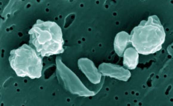
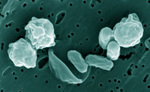
Bacillus odysseyi
Description:
Description: Colored micrograph of Bacillus odysseyi spores taken by a field-emission environmental SEM and magnified by 107. Spherical figures (about 2 µm diameter) are intact spores with exosporia. In center of image, spores (rod-shaped) were exposed to 0.5 mrad gamma radiation for 60 min, thus the exosporium (ribbon-shaped) separated. Source: http://www.kennislink.nl/web/show?id=134421. Author: James Kulleck, NASA/JPL.
Included On The Following Pages:
- Life (creatures)
- Cellular (cellular organisms)
- Bacteria
- Firmicutes (gram-positive bacteria)
- Bacilli
- Bacillales
- Bacillaceae
- Lysinibacillus
- Lysinibacillus odysseyi
This image is not featured in any collections.
Source Information
- license
- cc-publicdomain
- creator
- James Kulleck, NASA/JPL
- source
- http://www.kennislink.nl/web/show?id=134421
- original
- original media file
- visit source
- partner site
- Wikimedia Commons
- ID