Echinococcus vogeli HHS
Description:
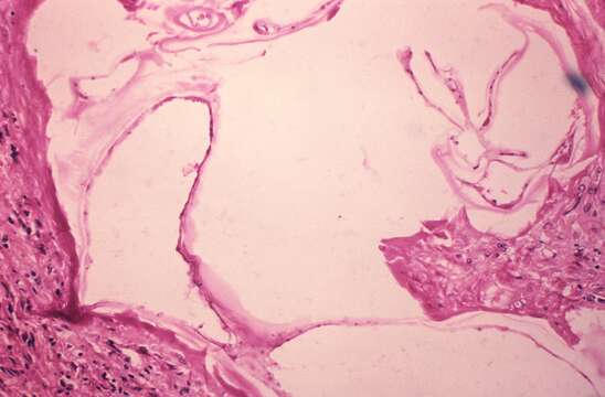
Description: English: This is a photomicrograph of a tissue sample harvested from a cyst, which was harbored inside a gorilla. In this view, the cyst interior revealed the presence of necrotic debris, and interspersed protoscolices of the microscopic tapeworm, Echinococcus vogeli. The larval stage of this microscopic tapeworm is one of the causative agents of alveolar hydatid disease (AHD), an infection in humans that causes parasitic tumors to form, mainly in the liver, but can also appear in other organs as well. Date: 1978. Source: : This media comes from the Centers for Disease Control and Prevention's Public Health Image Library (PHIL), with identification number #2895. Note: Not all PHIL images are public domain; be sure to check copyright status and credit authors and content providers. العربية | Deutsch | English | македонски | slovenščina | +/−. Author: Department of Health and Human Services.
Included On The Following Pages:
- Biota
- Eukaryota (eukaryotes)
- Unikonta
- Opisthokonta (opisthokonts)
- Distaplia
- Filozoa
- Apoikozoa
- Animalia
- Eumetazoa
- Bilateria
- Protostomia (protostomes)
- Platyzoa
- Platyhelminthes (flatworms)
- Cestoda (cestodes)
- Eucestoda
- Cyclophyllidea
- Taeniidae
- Echinococcus
- Echinococcus vogeli
This image is not featured in any collections.
Source Information
- license
- cc-publicdomain
- creator
- Department of Health and Human Services
- source
- {{CDC-PHIL|2895}}
- original
- original media file
- visit source
- partner site
- Wikimedia Commons
- ID