Chlamydomanas reinhardtii Flagella 9 - TEM
Description:
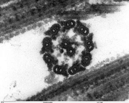
Description: Transmission electron microscope image, showing an example of green algae (Chlorophyta). Chlamydomanas reinhardtii is a unicellular flagellate used as a model system in molecular genetics work and flagellar motility studies. This image is a thin x-section cut through the isolated axoneme. Chlamydomonas flagella have the "9+2" structure characteristic of all eukaryotic cells. The axoneme has a central unit containing two single microtubules and nine peripheral doublet microtubules (known as the "9+2"). Dynein sidearms project from the A tubule of each doublet. Also visible in this image are the radial spokes and the inner sheath. Smith, E.F and P.A. Lefebvre (1996) "PF16 Encodes a Protein with Armadillo Repeats and Localizes to a Single Microtubule of the Central Apparatus in Chlamydomonas Flagella", J. Cell Biology, 132(3): 359-370 JEOL 100CX TEM. Source: http://remf.dartmouth.edu/imagesindex.html http://remf.dartmouth.edu/images/algaeTEM/source/14.html. Author: Elizabeth Smith, Louisa Howard, Erin Dymek. Permission(Reusing this file): PD.
Included On The Following Pages:
- Life
- Cellular
- Eukaryota
- Archaeplastida (plants)
- Chloroplastida
- Chlorophyta
- Chlorophyceae
- Chlamydomonadales
- Chlamydomonadaceae
- Chlamydomonas
- Chlamydomonas reinhardtii
This image is not featured in any collections.
Source Information
- license
- cc-publicdomain
- creator
- Elizabeth Smith, Louisa Howard, Erin Dymek
- original
- original media file
- visit source
- partner site
- Wikimedia Commons
- ID