197-Zika Virus-5ire glyc
Description:
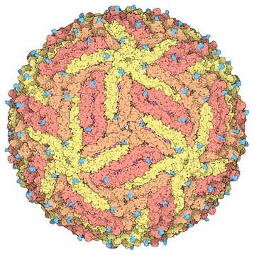
Description: English: Space-fill drawing of the Zika virus capsid, with the capsid proteins in shades of yellow and orange to show the icosahedral symmetry. The membrane proteins in the under layer (magenta) show through in some places, and the cyan protrusions are attached carbohydrate chains. Drawn by David Goodsell from the cryoEM structure 5ire. Date: 1 June 2016. Source: RCSB Molecule of the Month 197, June 2016. Author: David Goodwill.
Included On The Following Pages:
This image is not featured in any collections.
Source Information
- license
- cc-by-3.0
- copyright
- David Goodwill
- creator
- David Goodwill
- source
- RCSB Molecule of the Month 197, June 2016
- original
- original media file
- visit source
- partner site
- Wikimedia Commons
- ID