Taphrina deformans asci
Description:
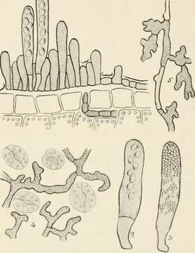
Description: Taphrina deformans. Asci at cuticula leaf. Fig. 31. I, Exoascus deformans, showing asci in various stages of development bursting through the cuticula of the leaf; 2, ascus of Exoascus pruni, showing stalk-cell at base of ascus, and eight spores; 3, ascus of Taphrina aurea filled with secondary spores produced by budding of the ascospores ; 4, surface view of mycelium of Taphrina sadebeckii on leaf of Alnus glutinosa ; 5, differentiation of fertile or ascogenous hyphae from vegetative hyphae of Taphrina sadebeckii. (Figs. 4 and 5 after Sadebeck.) All highly mag. upon. The asci at first contain eight spores, but in the majority of instances these spores germinate in the ascus, by the process of budding or germination, as in yeasts. Date: 1910. Source: https://www.flickr.com/photos/internetarchivebookimages/20966682015/in/photolist-rqbChX-pxZSbV-byE4od-4Qa9h6-hGtxDJ-fD7TtY-eiML2g-PUk5J-9GX75w-eiTsRJ-6yjVJr-eiTs25-eiTsqJ-4Wuboj-cqeTa1-nve8ZF-9a3bU1-ac8rFq-eiMKwM-nLRBzZ-BtNHcZ-oxXBsp-tDTo2B-oeGt83-4G7LDa-99Z4Mc-ziZUKt-rp5ZkQ-eiMK4t-GC18Ps-c8XRHo-ovZHz3-wDJXcS-xEjQ9S-xQgtkg-xAeicf-wVXFnc-xSNsWR-wVVvDD-wEjwf8-xBc66B-xthm9F-wxGqyg-wxycaE-oeeuqr-xT452i-xWKJ6v-ovw9xL-xzUsDF-xUuJBd. Author: Massee, George.
Included On The Following Pages:
- Life (creatures)
- Cellular (cellular organisms)
- Eukaryota (eukaryotes)
- Opisthokonta (opisthokonts)
- Nucletmycea
- Fungi (mushrooms, lichens, molds, yeasts and relatives)
- Dikarya
- Ascomycota (sac fungi)
- Taphrinomycetes
- Taphrinales
- Taphrinaceae
- Taphrina
- Taphrina deformans
This image is not featured in any collections.
Source Information
- license
- cc-publicdomain
- creator
- Massee, George
- source
- Flickr user ID internetarchivebookimages
- original
- original media file
- visit source
- partner site
- Wikimedia Commons
- ID