Hela cells with adenovirus
Description:
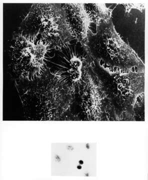
Summary.mw-parser-output table.commons-file-information-table,.mw-parser-output.fileinfotpl-type-information{border:1px solid #a2a9b1;background-color:#f8f9fa;padding:5px;font-size:95%;border-spacing:2px;box-sizing:border-box;margin:0;width:100%}.mw-parser-output table.commons-file-information-table>tbody>tr,.mw-parser-output.fileinfotpl-type-information>tbody>tr{vertical-align:top}.mw-parser-output table.commons-file-information-table>tbody>tr>td,.mw-parser-output table.commons-file-information-table>tbody>tr>th,.mw-parser-output.fileinfotpl-type-information>tbody>tr>td,.mw-parser-output.fileinfotpl-type-information>tbody>tr>th{padding:4px}.mw-parser-output.fileinfo-paramfield{background:#ccf;text-align:right;padding-right:0.4em;width:15%;font-weight:bold}.mw-parser-output.commons-file-information-table+table.commons-file-information-table,.mw-parser-output.commons-file-information-table+div.commons-file-information-table>table{border-top:0;padding-top:0;margin-top:-8px}@media only screen and (max-width:719px){.mw-parser-output table.commons-file-information-table,.mw-parser-output.commons-file-information-table.fileinfotpl-type-information{border-spacing:0;padding:0;word-break:break-word;width:100%!important}.mw-parser-output.commons-file-information-table>tbody,.mw-parser-output.fileinfotpl-type-information>tbody{display:block}.mw-parser-output.commons-file-information-table>tbody>tr>td,.mw-parser-output.commons-file-information-table>tbody>tr>th,.mw-parser-output.fileinfotpl-type-information>tbody>tr>td,.mw-parser-output.fileinfotpl-type-information>tbody>tr>th{padding:0.2em 0.4em;text-align:left;text-align:start}.mw-parser-output.commons-file-information-table>tbody>tr,.mw-parser-output.fileinfotpl-type-information>tbody>tr{display:flex;flex-direction:column}.mw-parser-output.commons-file-information-table+table.commons-file-information-table,.mw-parser-output.commons-file-information-table+div.commons-file-information-table>table{margin-top:-1px}.mw-parser-output.fileinfo-paramfield{box-sizing:border-box;flex:1 0 100%;width:100%}} Description: English: Title HeLa Cells with Adenovirus Description A scanning electron micrograph of cultured HeLa cells originally derived many years ago from a woman's cancerous cervical tissue. A light micrograph (x130) of the same cells (inset) reveals rounded double cells in the center in the process of dividing. This HeLa cell (named after patient Henrietta Lacks) has been infected with adenovirus. After 4-1/2 hours the HeLa cell's surface becomes rough. The multiple surface blebs on this cell characteristic for a certain stage of cell division that both normal and cancer cells go through. Research with the SEM has established the extraordinarily responsive nature of cell surface form. This instrument records, in pictures, specific cell reactions to various changes in the cells environment and maps the distribution of surface binding sites for biologically important molecules such as hormone, antigens, and pharmacologic agents. Topics/Categories Cells or Tissue, Abnormal Cells or Tissue Type B&W, Photo Source Dr. Timothy Triche. National Cancer Institute. Date: 1976. Source: : This image was released by the National Cancer Institute, an agency part of the National Institutes of Health, with the ID 1942 (image) (next). This tag does not indicate the copyright status of the attached work. A normal copyright tag is still required. See Commons:Licensing. Deutsch | English | français | македонски | +/−. Author: Unknown photographer. Permission(Reusing this file): Reuse Restrictions None - This image is in the public domain and can be freely reused. Please credit the source and/or author listed above.
Included On The Following Pages:
This image is not featured in any collections.
Source Information
- license
- cc-publicdomain
- source
- {{NCI Visuals Online|1942}}
- original
- original media file
- visit source
- partner site
- Wikimedia Commons
- ID