CroV reconstruction
Description:
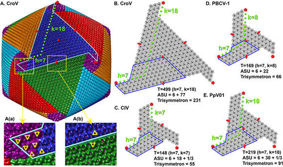
Description: English: Cryo-EM reconstruction of the CroV virion and capsomer arrangements of other giant icosahedral viruses. (A) Reconstruction of the CroV capsid. The isosurface of the map was colored by pentasymmetrons (purple) and trisymmetrons (blue, red, green, cyan and orange). One of the 30 edges of the icosahedron is marked by a cyan line. Two surface areas (a,b) are magnified and selected capsomers are labeled by yellow triangles to show their orientations. (B–E) Isolated icosahedral faces of CroV, PBCV-1, CIV and PpV01 capsids are shown schematically. Their T-numbers, asymmetric unit capsomer numbers, and trisymmetron capsomer numbers are listed. 5-fold, 3-fold, and 2-fold symbols are indicated in red and ASUs are outlined in blue. Date: 14 July 2017. Source: https://www.nature.com/articles/s41598-017-05824-w/figures/2. Author: Xiao, C., Fischer, M.G., Bolotaulo, D.M., Ulloa-Rondeau, N., Avila, G.A., and Suttle, C.A. (2017) Cryo-EM reconstruction of the Cafeteria roenbergensis virus capsid suggests novel assembly pathway for giant viruses. Scientific Reports 7: 5484. Other versions: .
.jpg)
Included On The Following Pages:
This image is not featured in any collections.
Source Information
- license
- cc-publicdomain
- creator
- Xiao, C., Fischer, M.G., Bolotaulo, D.M., Ulloa-Rondeau, N., Avila, G.A., and Suttle, C.A. (2017) Cryo-EM reconstruction of the Cafeteria roenbergensis virus capsid suggests novel assembly pathway for giant viruses. Scientific Reports 7: 5484.
- source
- https://www.nature.com/articles/s41598-017-05824-w/figures/2
- original
- original media file
- visit source
- partner site
- Wikimedia Commons
- ID