CroV TEM
Description:
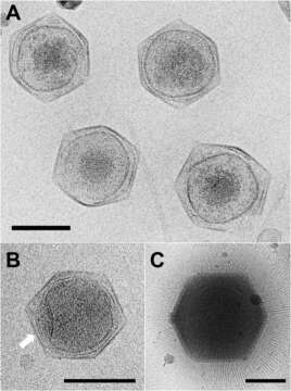
Description: English: Cryo-EM images of CroV compared to APMV. (A) Cryo-electron micrograph of four CroV particles. (B) Single CroV particle with concave core depression (white arrow). (C) Single APMV particle. Scale bars in (A–C) represent 2,000 Å. Date: 14 July 2017. Source: https://www.nature.com/articles/s41598-017-05824-w/figures/1. Author: Xiao, C., Fischer, M.G., Bolotaulo, D.M., Ulloa-Rondeau, N., Avila, G.A., and Suttle, C.A. (2017) Cryo-EM reconstruction of the Cafeteria roenbergensis virus capsid suggests novel assembly pathway for giant viruses. Scientific Reports 7: 5484. Other versions: .
.jpg)
Included On The Following Pages:
This image is not featured in any collections.
Source Information
- license
- cc-publicdomain
- creator
- Xiao, C., Fischer, M.G., Bolotaulo, D.M., Ulloa-Rondeau, N., Avila, G.A., and Suttle, C.A. (2017) Cryo-EM reconstruction of the Cafeteria roenbergensis virus capsid suggests novel assembly pathway for giant viruses. Scientific Reports 7: 5484.
- source
- https://www.nature.com/articles/s41598-017-05824-w/figures/1
- original
- original media file
- visit source
- partner site
- Wikimedia Commons
- ID