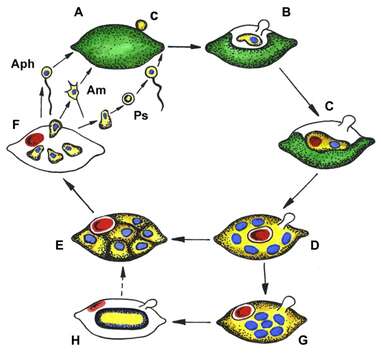
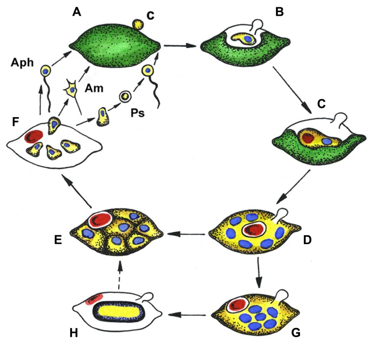
Generalized life cycle of aphelids.
Description:
Description: English: Aphelidium (Aph), Amoeboaphelidium (Am) and Pseudaphelidium (Ps), distinguished by zoospore structure and development. (A) Zoospore encystment (c) on the host surface, (B) propagule penetration into the host, (C) trophic amoeba with nucleus (blue) and residual body (red) engulfs host cytoplasm, (D) multinuclear plasmodium (yellow) totally replaced the host, contains several nuclei (blue) and central vacuole with residual body (red), (E) plasmodium divides producing uninuclear cells, (F) mature zoospores released from the empty host cell, (G) precursory stage to the resting spore with nuclei in the center, (H) resting spore with ejected residual body. Dotted line shows conceivable way from resting spore to divided plasmodium. Colors: green, host (alga) cytoplasm; yellow, parasitoid cytoplasm; blue, nucleus; red, residual body. Date: 28 March 2014. Source: http://journal.frontiersin.org/Journal/10.3389/fmicb.2014.00112/abstract. Author: Sergey A. Karpov, Maria A. Mamkaeva, Vladimir V. Aleoshin, Elena Nassonova Osu Lilje and Frank H. Gleason.
Included On The Following Pages:
- Life (creatures)
- Cellular (cellular organisms)
- Eukaryota (eukaryotes)
- Opisthokonta (opisthokonts)
- Nucletmycea
- Fungi (mushrooms, lichens, molds, yeasts and relatives)
- Aphelida
This image is not featured in any collections.
Source Information
- license
- cc-by-3.0
- copyright
- Sergey A. Karpov, Maria A. Mamkaeva, Vladimir V. Aleoshin, Elena Nassonova Osu Lilje and Frank H. Gleason.
- creator
- Sergey A. Karpov, Maria A. Mamkaeva, Vladimir V. Aleoshin, Elena Nassonova Osu Lilje and Frank H. Gleason.
- source
- http://journal.frontiersin.org/Journal/10.3389/fmicb.2014.00112/abstract
- original
- original media file
- visit source
- partner site
- Wikimedia Commons
- ID