Fmicb-07-02140-g002
Description:
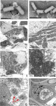
Description: English: Electron micrograph of infected rice leaves. Negative-stained sap of infected rice leaves (A,B). Ultrathin sections of infected rice leaves showing RSMV virions and cytopathological structures (C–G), the enlarged CS of (E) is shown in (F). The uninfected rice leaves cells are shown in (H). Black arrows indicate the RSMV virions with enveloped, red arrows indicate the vesicle structure with gathered virions in it. CW = cell wall, CH = chloroplast, CS = crystalline structure, M = mitochondrion, N = nucleus, V = Virion, and Vp = viroplasm. Date: 4 January 2017. Source: https://www.frontiersin.org/articles/10.3389/fmicb.2016.02140/full. Author: Xin Yang, Jilei Huang, Chuanhe Liu, Biao Chen, Tong Zhang, and Guohui Zhou.
Included On The Following Pages:
This image is not featured in any collections.
Source Information
- license
- cc-by-3.0
- copyright
- Xin Yang, Jilei Huang, Chuanhe Liu, Biao Chen, Tong Zhang, and Guohui Zhou
- creator
- Xin Yang, Jilei Huang, Chuanhe Liu, Biao Chen, Tong Zhang, and Guohui Zhou
- source
- https://www.frontiersin.org/articles/10.3389/fmicb.2016.02140/full
- original
- original media file
- visit source
- partner site
- Wikimedia Commons
- ID