Schistosomiasis haematobia
Description:
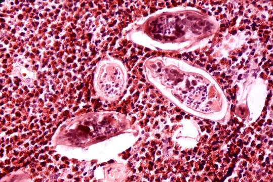
Description: English: Histopathology of schistosomiasis haematobia, bladder Histopathology of bladder shows eggs of Schistosoma haematobium surrounded by intense infiltrates of eosinophils. Parasite. Date: 1973. Source: : This media comes from the Centers for Disease Control and Prevention's Public Health Image Library (PHIL), with identification number #35. Note: Not all PHIL images are public domain; be sure to check copyright status and credit authors and content providers. العربية | Deutsch | English | македонски | slovenščina | +/−. Author: Photo Credit: Content Providers(s): CDC/ Dr. Edwin P. Ewing, Jr. Permission(Reusing this file): PD-USGov-HHS-CDC English: None - This image is in the public domain and thus free of any copyright restrictions. As a matter of courtesy we request that the content provider be credited and notified in any public or private usage of this image.
Included On The Following Pages:
- Biota
- Eukaryota (eukaryotes)
- Unikonta
- Opisthokonta (opisthokonts)
- Distaplia
- Filozoa
- Apoikozoa
- Animalia
- Eumetazoa
- Bilateria
- Protostomia (protostomes)
- Platyzoa
- Platyhelminthes (flatworms)
- Trematoda (flukes)
- Digenea (Parasitic Flatworms)
- Strigeatida
- Schistosomatidae (blood flukes)
- Mictosoma
- Schistosoma haematobium
This image is not featured in any collections.
Source Information
- license
- cc-publicdomain
- original
- original media file
- visit source
- partner site
- Wikimedia Commons
- ID