Algal Golgi body, 3D reconstruction (30466939665)
Description:
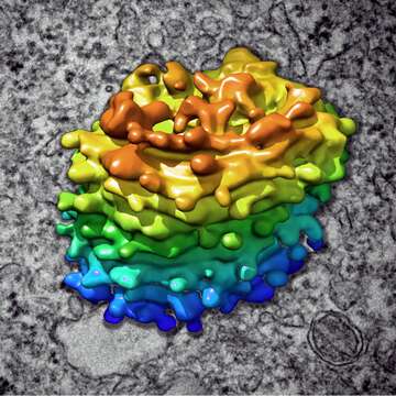
Description: 3D reconstruction of Chlamydomonas Golgi body, based on ZEISS Crossbeam FIB-SEM ion milling raw data. Blue/green: cis-golgi, yellow/orange: trans-golgi. www.zeiss.com/crossbeam Courtesy of Louise Hughes, Oxford Brookes University, UK. Images donated as part of a GLAM collaboration with Carl Zeiss Microscopy - please contact Andy Mabbett for details. Date: 20 October 2016, 10:37. Source: Algal Golgi body, 3D reconstruction. Author: ZEISS Microscopy from Germany.
Included On The Following Pages:
- Life (creatures)
- Cellular (cellular organisms)
- Eukaryota (eukaryotes)
- Archaeplastida (plants)
- Chloroplastida (green plants)
- Chlorophyta (chlorophytes)
- Chlorophyceae
- Chlamydomonadales
- Chlamydomonadaceae
- Chlamydomonas
This image is not featured in any collections.
Source Information
- license
- cc-by-3.0
- copyright
- ZEISS Microscopy
- creator
- ZEISS Microscopy
- source
- Flickr user ID zeissmicro
- original
- original media file
- visit source
- partner site
- Wikimedia Commons
- ID
.jpg)
.jpg)