Fmars-08-742498-g003
Description:
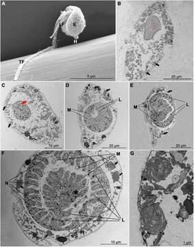
Summary.mw-parser-output table.commons-file-information-table,.mw-parser-output.fileinfotpl-type-information{border:1px solid #a2a9b1;background-color:#f8f9fa;padding:5px;font-size:95%;border-spacing:2px;box-sizing:border-box;margin:0;width:100%}.mw-parser-output table.commons-file-information-table>tbody>tr,.mw-parser-output.fileinfotpl-type-information>tbody>tr{vertical-align:top}.mw-parser-output table.commons-file-information-table>tbody>tr>td,.mw-parser-output table.commons-file-information-table>tbody>tr>th,.mw-parser-output.fileinfotpl-type-information>tbody>tr>td,.mw-parser-output.fileinfotpl-type-information>tbody>tr>th{padding:4px}.mw-parser-output.fileinfo-paramfield{background:#ccf;text-align:right;padding-right:0.4em;width:15%;font-weight:bold}.mw-parser-output.commons-file-information-table+table.commons-file-information-table,.mw-parser-output.commons-file-information-table+div.commons-file-information-table>table{border-top:0;padding-top:0;margin-top:-8px}@media only screen and (max-width:719px){.mw-parser-output table.commons-file-information-table,.mw-parser-output.commons-file-information-table.fileinfotpl-type-information{border-spacing:0;padding:0;word-break:break-word;width:100%!important}.mw-parser-output.commons-file-information-table>tbody,.mw-parser-output.fileinfotpl-type-information>tbody{display:block}.mw-parser-output.commons-file-information-table>tbody>tr>td,.mw-parser-output.commons-file-information-table>tbody>tr>th,.mw-parser-output.fileinfotpl-type-information>tbody>tr>td,.mw-parser-output.fileinfotpl-type-information>tbody>tr>th{padding:0.2em 0.4em;text-align:left;text-align:start}.mw-parser-output.commons-file-information-table>tbody>tr,.mw-parser-output.fileinfotpl-type-information>tbody>tr{display:flex;flex-direction:column}.mw-parser-output.commons-file-information-table+table.commons-file-information-table,.mw-parser-output.commons-file-information-table+div.commons-file-information-table>table{margin-top:-1px}.mw-parser-output.fileinfo-paramfield{box-sizing:border-box;flex:1 0 100%;width:100%}} Description: English: Electron micrographs of free-swimming dinospores and Amoebophrya sp. within Akashiwo sanguinea.(A) Scanning electron microscope image of Amoebophrya sp. dinospore showing the episome (E), hyposome (H), longitudinal flagellum (LF), transverse flagellum (TF), and flagellar hairs (black arrow).(B–G) Transmission electron microscope images of A. sanguinea infected with Amoebophrya sp. Black arrows indicated the chloroplast of A. sanguinea.(B) Uninfected A. sanguinea showing nucleus, nucleolus (red circle) and nucleus membrane.(C) 12 h infection with multiple parasite (red arrow) in host nucleus.(D) 30 h infection showing nascent mastigocoel (M) and multilobed cavity (L).(E) 54 h infection showing “beehive” with fully developed mastigocoel (M) and multilobed cavity (L).(F) Transverse section through 54 h Amoebophrya sp. trophont within the A. sanguinea nucleus. The host cytoplasm (H) was relegated by the parasite to the margin of the cell. The mastigocoel (M) contains numerous flagella (F), and the nucleus (N) line up in a continuous fashion along the folds of the “beehive.” Asterisk indicated the clusters of parasite trichocysts.(G) Detail of chloroplasts in the 54 h infected A. sanguinea. Date: 26 November 2021. Source: Chloroplast Ultrastructure and Photosynthetic Response of the Dinoflagellate Akashiwo sanguinea Throughout Infection by Amoebophrya sp. In: Frontiers in Marine Science, Volume 8 - 2021; Sec. Marine Ecosystem Ecology; doi:10.3389/fmars.2021.742498. Author: Tiantian Chen, Yun Liu, Zhangxi Hu, Shuqun Song, Caiwen Li. Other versions: This file has an extracted image: Fmars-08-742498-g003A.jpg..
Included On The Following Pages:
- Life (creatures)
- Cellular (cellular organisms)
- Eukaryota (eukaryotes)
- SAR (Stramenopiles, Alveolates, Rhizaria)
- Alveolata (alveolates)
- Dinophyceae
This image is not featured in any collections.
Source Information
- license
- cc-by-sa-3.0
- copyright
- Tiantian Chen, Yun Liu, Zhangxi Hu, Shuqun Song, Caiwen Li
- creator
- Tiantian Chen, Yun Liu, Zhangxi Hu, Shuqun Song, Caiwen Li
- source
- Chloroplast Ultrastructure and Photosynthetic Response of the Dinoflagellate Akashiwo sanguinea Throughout Infection by Amoebophrya sp. In: Frontiers in Marine Science, Volume 8 - 2021; Sec. Marine Ecosystem Ecology; doi:10.3389/fmars.2021.742498
- original
- original media file
- visit source
- partner site
- Wikimedia Commons
- ID