Fmars-08-720424-g002
Description:
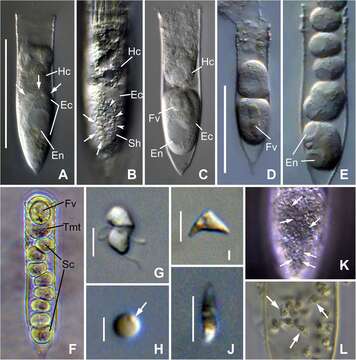
Summary.mw-parser-output table.commons-file-information-table,.mw-parser-output.fileinfotpl-type-information{border:1px solid #a2a9b1;background-color:#f8f9fa;padding:5px;font-size:95%;border-spacing:2px;box-sizing:border-box;margin:0;width:100%}.mw-parser-output table.commons-file-information-table>tbody>tr,.mw-parser-output.fileinfotpl-type-information>tbody>tr{vertical-align:top}.mw-parser-output table.commons-file-information-table>tbody>tr>td,.mw-parser-output table.commons-file-information-table>tbody>tr>th,.mw-parser-output.fileinfotpl-type-information>tbody>tr>td,.mw-parser-output.fileinfotpl-type-information>tbody>tr>th{padding:4px}.mw-parser-output.fileinfo-paramfield{background:#ccf;text-align:right;padding-right:0.4em;width:15%;font-weight:bold}.mw-parser-output.commons-file-information-table+table.commons-file-information-table,.mw-parser-output.commons-file-information-table+div.commons-file-information-table>table{border-top:0;padding-top:0;margin-top:-8px}@media only screen and (max-width:719px){.mw-parser-output table.commons-file-information-table,.mw-parser-output.commons-file-information-table.fileinfotpl-type-information{border-spacing:0;padding:0;word-break:break-word;width:100%!important}.mw-parser-output.commons-file-information-table>tbody,.mw-parser-output.fileinfotpl-type-information>tbody{display:block}.mw-parser-output.commons-file-information-table>tbody>tr>td,.mw-parser-output.commons-file-information-table>tbody>tr>th,.mw-parser-output.fileinfotpl-type-information>tbody>tr>td,.mw-parser-output.fileinfotpl-type-information>tbody>tr>th{padding:0.2em 0.4em;text-align:left;text-align:start}.mw-parser-output.commons-file-information-table>tbody>tr,.mw-parser-output.fileinfotpl-type-information>tbody>tr{display:flex;flex-direction:column}.mw-parser-output.commons-file-information-table+table.commons-file-information-table,.mw-parser-output.commons-file-information-table+div.commons-file-information-table>table{margin-top:-1px}.mw-parser-output.fileinfo-paramfield{box-sizing:border-box;flex:1 0 100%;width:100%}} Description: English: Photomicrograph of Euduboscquella triangula n. sp. from live (A–J), Lugol-fixed (K), and Bouin-fixed (L) specimens.(A) Lorica containing infected host cell with a late trophont of E. triangula (arrows indicate the margins of parasite cytoplasm).(B) Trophont showing shield margins (arrowheads) with intertwining grooves (arrows).(C) Tomont emerged from host with food vacuole.(D,E) Sporogenic division.(F) One tomont and eight sporocytes resulting from different trophonts of a double infected host.(G) Mushroom-shaped dinospore.(H) Non-motile spherical spore with granule on cell surface (arrow).(I,J) Lateral (I) and apical (J) view of non-motile triangular-shaped spores.(K) Shield with intertwining grooves (arrows) in Lugol-fixed specimen.(L) Triangular-shaped spores (arrows) in host lorica from field sample fixed with Bouin immediately after collection.Ec, Euduboscquella cytoplasm; En, Euduboscquella nucleus; Fv, food vacuole; Hc, Helicostomella longa cytoplasm; Sc, sporocytes; Sh, shield; Tmt, tomont.Scale bar: (A,D), 50 μm; (G,I,J), 5 μm; (H), 2 μm. Date: 12 August 2021. Source: A Novel Parasitic, Syndinean Dinoflagellate Euduboscquella triangula Infecting the Tintinnid Helicostomella longa. In: Frontiers in Marine Science, Volume 8 - 2021, Sec. Aquatic Microbiology, doi:10.3389/fmars.2021.720424. Author: Jung Min Choi, Jae Ho Jung, Ki Hong Kim, D. Wayne Coats, Young Ok Kim.
Included On The Following Pages:
- Life (creatures)
- Cellular (cellular organisms)
- Eukaryota (eukaryotes)
- SAR (Stramenopiles, Alveolates, Rhizaria)
- Alveolata (alveolates)
- Dinophyceae
This image is not featured in any collections.
Source Information
- license
- cc-by-sa-3.0
- copyright
- Jung Min Choi, Jae Ho Jung, Ki Hong Kim, D. Wayne Coats, Young Ok Kim
- creator
- Jung Min Choi, Jae Ho Jung, Ki Hong Kim, D. Wayne Coats, Young Ok Kim
- source
- A Novel Parasitic, Syndinean Dinoflagellate Euduboscquella triangula Infecting the Tintinnid Helicostomella longa. In: Frontiers in Marine Science, Volume 8 - 2021, Sec. Aquatic Microbiology, doi:10.3389/fmars.2021.720424.
- original
- original media file
- visit source
- partner site
- Wikimedia Commons
- ID