Brookesia nana female anatomy
Description:
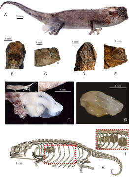
Description: English: Morphological characters of Brookesia nana sp. nov.: (A) preserved holotype (ZSM 1660/2012) in lateral view, showing right everted hemipenis, (B) head in dorsal and (C) lateral (mirrored, indicated with asterisk) views; head of female paratype (UADBA-R/FGZC 3752) in (D) dorsal and (E) lateral views; (F, G) close-ups of everted left hemipenis of holotype photographed under different light conditions; (H) micro-CT scan image of the female paratype in lateral view showing its skeleton. The inset image (I) shows the area marked by the stippled square viewed at a different rendering threshold, showing two developing eggs in the females’ ovaries. Date: 28 January 2021. Source: Glaw, F., Köhler, J., Hawlitschek, O. et al. Extreme miniaturization of a new amniote vertebrate and insights into the evolution of genital size in chameleons. Sciientific Reports 11, 2522 (2021).doi:10.1038/s41598-020-80955-1. Author: Frank Glaw, Jörn Köhler, Oliver Hawlitschek, Fanomezana M. Ratsoavina, Andolalao Rakotoarison, Mark D. Scherz & Miguel Vences.
Included On The Following Pages:
- Biota
- Eukaryota (eukaryotes)
- Unikonta
- Opisthokonta (opisthokonts)
- Distaplia
- Filozoa
- Apoikozoa
- Animalia
- Eumetazoa
- Bilateria
- Deuterostomia (deuterostomes)
- Chordata (Chordates)
- Olfactores
- Craniata
- Vertebrata (vertebrates)
- Gnathostomata (jawed fish)
- Osteichthyes
- Sarcopterygii (Lobe-finned fishes)
- Dipnotetrapodomorpha
- Tetrapodomorpha
- Tetrapoda (terrestrial vertebrates)
- Reptiliomorpha
- Amniota (amniotes)
- Sauropsida
- Reptilia (Reptiles)
- Eureptilia
- Romeriida
- Diapsida (diapsid)
- Neodiapsida
- Sauria
- Lepidosauromorpha
- Lepidosauria (lepidosaur)
- Squamata (lizards and snakes)
- Episquamata
- Toxicofera
- Iguania
- Chamaeleonidae (chameleons)
- Brookesiinae
- Brookesia (Leaf chameleon)
- Brookesia nana
This image is not featured in any collections.
Source Information
- license
- cc-by-3.0
- copyright
- Frank Glaw, Jörn Köhler, Oliver Hawlitschek, Fanomezana M. Ratsoavina, Andolalao Rakotoarison, Mark D. Scherz & Miguel Vences
- creator
- Frank Glaw, Jörn Köhler, Oliver Hawlitschek, Fanomezana M. Ratsoavina, Andolalao Rakotoarison, Mark D. Scherz & Miguel Vences
- source
- Glaw, F., Köhler, J., Hawlitschek, O. et al. Extreme miniaturization of a new amniote vertebrate and insights into the evolution of genital size in chameleons. Sciientific Reports 11, 2522 (2021).doi:10.1038/s41598-020-80955-1
- original
- original media file
- visit source
- partner site
- Wikimedia Commons
- ID