Image of Manota
Description:
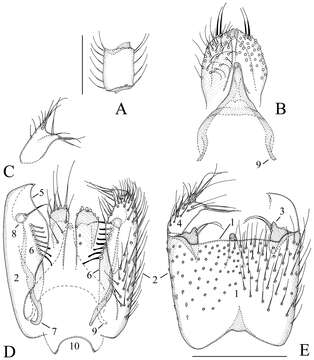
Figure 3.Manota belalongensis sp. n. (holotype). A Antennal flagellomere 4, lateral view B Aedeagus and hypoproct. ventral view C Gonostylus, dorsal view D Hypopygium, dorsal view E Hypopygium, ventral view. Scale 0.1 mm. 1 = sternite 9, 2 = gonocoxa, 3 = parastylar lobe, 4 = gonostylus, 5 = dorsal mesial margin of gonocoxa, 6 = plate-like lobe ventrally from the dorsal mesial margin of gonocoxa, 7 = gonocoxal apodeme, 8 = juxtagonostylar seta, 9 = aedeagal apodeme, 10 = tergite 9.
Included On The Following Pages:
- Life
- Cellular
- Eukaryota (eukaryotes)
- Opisthokonta (opisthokonts)
- Metazoa (animals)
- Bilateria
- Protostomia (protostomes)
- Ecdysozoa (ecdysozoans)
- Arthropoda (arthropods)
- Pancrustacea
- Hexapoda (hexapods)
- Insecta (insects)
- Pterygota (winged insects)
- Neoptera (neopteran)
- Endopterygota (endopterygotes)
- Diptera (flies)
- Bibionomorpha
- Mycetophilidae (fungus gnats)
- Manota
- Manota belalongensis
- Panarthropoda
This image is not featured in any collections.
Source Information
- license
- cc-by-3.0
- copyright
- Jan Ševčík, Heikki Hippa, Rodzay Abdul Wahab
- bibliographic citation
- Ševčík J, Hippa H, Wahab R (2014) Diversity of Manota Williston (Diptera, Mycetophilidae) in Ulu Temburong National Park, Brunei ZooKeys 428: 57–77
- original
- original media file
- visit source
- partner site
- Zookeys
- ID