Image of Echinoderes hwiizaa Yamasaki & Fujimoto 2014
Description:
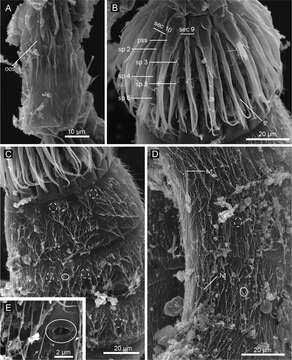
Figure 9.Echinoderes hwiizaa sp. n., scanning electron micrographs. A Mouth cone B introvert, lateral view C neck and segments1–3, lateral view D segments 5–7, lateral view E close up of laterodorsal type 2 glandular cell outlet on segment 2. Complete circles indicate type 2 glandular cell outlet; dashed circles indicate sensory spots. Abbreviations: lvt, lateroventral tubule; oos, outer oral style; pss, primary spinoscalid; sec, introvert sector followed by sector number; sp, spinoscalid followed by ring number.
Included On The Following Pages:
- Life (creatures)
- Cellular (cellular organisms)
- Eukaryota (eukaryotes)
- Opisthokonta (opisthokonts)
- Metazoa (Animal)
- Bilateria
- Protostomia (protostomes)
- Ecdysozoa (ecdysozoans)
- Kinorhyncha (mud dragons)
- Cyclorhagida
- Echinoderidae
- Echinoderes
- Echinoderes hwiizaa
- Scalidophora
This image is not featured in any collections.
Source Information
- license
- cc-by-3.0
- copyright
- Hiroshi Yamasaki, Shinta Fujimoto
- bibliographic citation
- Yamasaki H, Fujimoto S (2014) Two new species in the Echinoderes coulli group (Echinoderidae, Cyclorhagida, Kinorhyncha) from the Ryukyu Islands, Japan ZooKeys 382: 27–52
- original
- original media file
- visit source
- partner site
- Zookeys
- ID