Image of wafer-lid trapdoor spiders
Description:
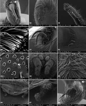
Figures 57–68.Scanning electron micrographs of Aptostichus atomarius specimens (female MY3432, 57–67; male MY2979 68). 57 left chelicerae, anterior ventral aspect 58 left pedipalp claw, retrolateral aspect 59 leg I, tarsal claw, retrolateral aspect 60 tarsal scopulae, leg I 61 leg IV tarsal claws, retrolateral aspect 62 leg IV preening comb, tarsus/metatarsus junction 63 leg I, tarsus, dorsal aspect, tarsal organ 64 spinnerets 65 spinning field, distal aspect of PLS 66, 67 cleared spermathecae and close-up of left lobe 68 pedipalp, retrolateral aspect, arrow indicates position of spines on cymbium.
Included On The Following Pages:
- Life (biota)
- Cellular
- Eukaryota (eukaryotes)
- Opisthokonta (opisthokonts)
- Metazoa (animals)
- Bilateria (bilaterians)
- Protostomia (protostomes)
- Ecdysozoa (ecdysozoans)
- Arthropoda (arthropods)
- Chelicerata (Chelicerates)
- Arachnida (arachnids)
- Araneae (spiders)
- Opisthothelae
- Mygalomorphae (mygalomorphs)
- Euctenizidae (wafer-lid trapdoor spiders)
- Aptostichus
- Panarthropoda
This image is not featured in any collections.
Source Information
- license
- cc-by-3.0
- copyright
- Jason E. Bond
- bibliographic citation
- Bond J (2012) Phylogenetic treatment and taxonomic revision of the trapdoor spider genus Aptostichus Simon (Araneae, Mygalomorphae, Euctenizidae) ZooKeys 252: 1–209
- original
- original media file
- visit source
- partner site
- Zookeys
- ID