in vivo
Description:
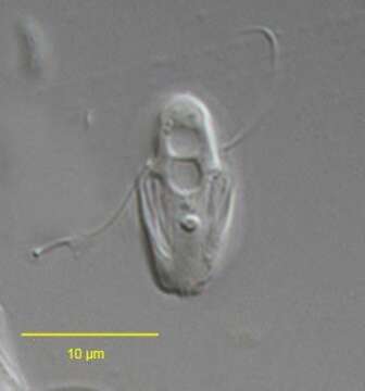
Trepomonas agilis (Dujardin, 1841). Cell is ovoid, but S-shaped in cross section. Two nuclei are located anteriorly. Two groups of flagella are inserted laterally at the end of each groove: two long flagella and six short flagella. The short falgella are less than half the cell length and lie in the grooves (seen best here to viewer's left). Contractile vacuoles are seen.Collected from putifying sample from a freshwater pond near Boise, Idaho.DIC.
Included On The Following Pages:
- Life (creatures)
- Cellular (cellular organisms)
- Eukaryota (eukaryotes)
- Excavates (excavates)
- Metamonada (metamonad)
- Fornicata
- Diplomonadida
- Hexamitidae
- Hexamitinae
- Trepomonas
- Trepomonas agilis
This image is not featured in any collections.
Source Information
- license
- cc-by-nc
- author
- William Bourland
- provider
- micro*scope
- original
- original media file
- visit source
- partner site
- micro*scope
- ID