Oral infraciliature
Description:
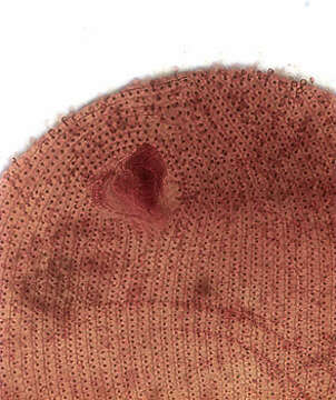
Oral infraciliature (ventral view) of Epenardia myriophylli (Penard, 1922) Corliss, 1971, a large glaucomid ciliate. This cell is slighlty compressed. The body is ellipsoid with minimal dorsoventral flattening. The relatively small obliquely oriented cytostome is in the anterior 1/4. The three adoral membranelles are located in the deep buccal cavity. There is an undulating membrane on the right. E. myriophylli differs from Glaucoma species by its dense (80-110) somatic kineties and the broad M3 which is wider than M2 and a preoral suture in the long axis. There are approximately 9 postoral kineties. The spherical macronucleus and micronucleus are centrally located. The contractile vacuole empties through a single excretory pore on the right dorsal surface. The pellicle is pitted with regular square depressions. Collected from a freshwater drainage ditch near Boise, Idaho. April 2005.Silver carbonate stain (see Foissner, W. Europ. J. Protistol., 27:313-330;1991). Brightfield.
Included On The Following Pages:
- Life (creatures)
- Cellular (cellular organisms)
- Eukaryota (eukaryotes)
- SAR (Stramenopiles, Alveolates, Rhizaria)
- Alveolata (alveolates)
- Ciliophora (ciliates)
- Intramacronucleata
- Oligohymenophorea
- Hymenostomatida
- Tetrahymenina
- Glaucomidae
- Epenardia
- Epenardia myriophylli
This image is not featured in any collections.
Source Information
- license
- cc-by-nc
- author
- William Bourland
- provider
- micro*scope
- original
- original media file
- visit source
- partner site
- micro*scope
- ID