Infraciliature
Description:
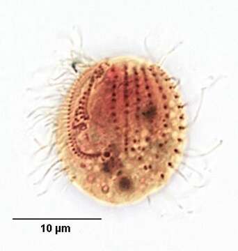
Ventral view of the small scutociliate, Cyclidium glaucoma (Mueller, 1773). The infraciliature has been stained by the silver carbonate technique (see Foissner, W. Europ. J. Protistol.27,313-330;1991). 15-30 microns in length. The cell is ovoid and laterally compressed. A prominent paraoral membrane borders the peristome on the right, curving around it posteriorly to form a pouch. The paraoral membrane extends at least 1/2 the cell length. Just to the left of the paraoral membrane three small polykinetids correspond to adoral membranelles 1, 2 and 3. The nonciliated basal bodies of the scuticovestige at the posterior end of the paraoral membrane are visible in this specimen. Longitudinal somatic kineties are visible to the left of the cytostome. Collected from stagnant organically enriched freshwater near Boise, Idaho August 2004. Brightfield
Included On The Following Pages:
- Life (creatures)
- Cellular (cellular organisms)
- Eukaryota (eukaryotes)
- SAR (Stramenopiles, Alveolates, Rhizaria)
- Alveolata (alveolates)
- Ciliophora (ciliates)
- Intramacronucleata
- Oligohymenophorea
- Scuticociliatia
- Pleuronematida
- Cyclidiidae
- Cyclidium
- Cyclidium glaucoma
This image is not featured in any collections.
Source Information
- license
- cc-by-nc
- author
- William Bourland
- provider
- micro*scope
- original
- original media file
- visit source
- partner site
- micro*scope
- ID