Oral infraciliature
Description:
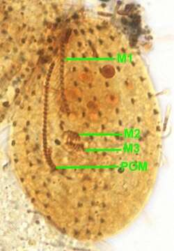
Ventral infraciliature of the pleuronematine scuticociliate, Ctedoctema acanthocryptum (Stokes, 1884). Small elongate to oval ciliate that is slightly dorsoventrally compressed. There is a prominent paraoral membrane (POM), which begins subapically and extends approximately two-third down the length of the cell on the right side of a shallow groove. The cytostome is at the posterior end of this groove. There is a three part adoral zone of membranelles on the left of the peristome. M1 is a longitudinal array of dikinetids from the sub-apical pole to about midway down the cell. The posterior end of M1 is obscured here by the densely stained macronucleus. M2 and M3 are small, oblique patches of kinetids posterior to M1. Somatic ciliation uniform. Ovoid macronucleus in anterior body half. Collected from freshwater pond near Boise, Idaho October 2004. Silver carbonate stain (see Foissner, W.Europ. J. Protistol.27, 313-330; 1991). Brightfield optics.
Included On The Following Pages:
- Life (creatures)
- Cellular (cellular organisms)
- Eukaryota (eukaryotes)
- SAR (Stramenopiles, Alveolates, Rhizaria)
- Alveolata (alveolates)
- Ciliophora (ciliates)
- Intramacronucleata
- Oligohymenophorea
- Scuticociliatia
- Pleuronematida
- Ctedoctematidae
- Ctedoctema
- Ctedoctema acanthocryptum
This image is not featured in any collections.
Source Information
- license
- cc-by-nc
- author
- William Bourland
- provider
- micro*scope
- original
- original media file
- visit source
- partner site
- micro*scope
- ID