Portrait
Description:
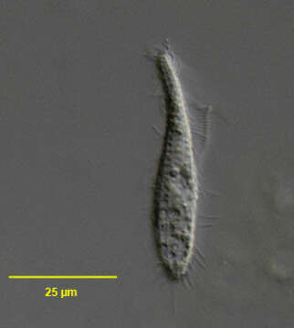
Portrait of the hymenostome ciliate, Cohnilembus verminus (Muller, 1786, Kahl, 1933). C. vermiformis, described in Careyâs key to species of Cohnilembus (Carey, P.G., Marine Interstitial Ciliates. P. 136. Chapman and Hall, London,1992), is probably a subjective synonym. The cell outline is a teardrop shape with a slender neck region extending about ½ cell length. The peristome area extends from the anterior end posteriorly to about mid-body. The cytostome is at the posterior end of the peristome. There are 2 membranelles running along the right edge if of the peristome. The more prominent one (seen here) protrudes, flag-like, perpendicular to the long axis. Longitudinal somatic kineties (9 or 100 are uniformly spaced. There is a single long caudal cilium present. The single contractile vacuole is at the posterior terminus. There is a single central macronucleus and adjacent micronucleus (not well-seen here). Collected from a commercial saltwater aquarium in Boise,Idaho August 2004. DIC optics.
Included On The Following Pages:
- Life (creatures)
- Cellular (cellular organisms)
- Eukaryota (eukaryotes)
- SAR (Stramenopiles, Alveolates, Rhizaria)
- Alveolata (alveolates)
- Ciliophora (ciliates)
- Intramacronucleata
- Oligohymenophorea
- Scuticociliatia
- Philasterida
- Cohnilembidae
- Cohnilembus
- Cohnilembus verminus
This image is not featured in any collections.
Source Information
- license
- cc-by-nc
- author
- Bill Bourland
- provider
- micro*scope
- original
- original media file
- visit source
- partner site
- micro*scope
- ID