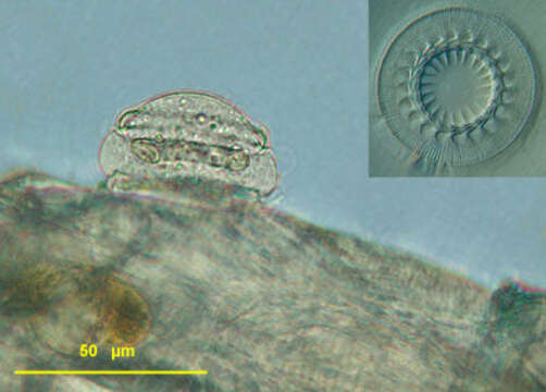
portrait & adhesive disk
Description:
Description: Lateral view of the motile peritrich ciliate. Specimens were found on Eudiaptomus vulgaris, a common copepod. Compared with the bobbin-shaped Trichodina pediculus the lateral view of these cells is round, more dome-shaped, the size varying from 42 to 52 µm. The macronucleus is found in the transverse axis of the cell, almost circular. The large micronucleus lies at the outside of the c-shaped macronucleus. The conspicuous adhesive disk is located at the aboral side of the cell and, functioning as a holdfast organelle, attaches the ciliate to the surface of itâs host. The anatomy of the adhesive disk differs between species of the genus. The picture in the upper right angle shows an adhesive disk separated from itâs cell, counting 20 denticles
Included On The Following Pages:
- Life (creatures)
- Cellular (cellular organisms)
- Eukaryota (eukaryotes)
- SAR (Stramenopiles, Alveolates, Rhizaria)
- Alveolata (alveolates)
- Ciliophora (ciliates)
- Intramacronucleata
- Oligohymenophorea
- Peritrichia
- Mobilida
- Trichodinidae
- Trichodina
- Trichodina domerguei
- Trichodina domerguei megamicronucleata
This image is not featured in any collections.
Source Information
- license
- cc-by-nc
- author
- Dr. Dr. Josef Brief
- provider
- micro*scope
- original
- original media file
- visit source
- partner site
- micro*scope
- ID