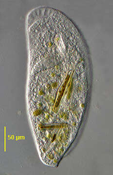
Dorsal view
Description:
Dorsal view of the large Chlamydodontid ciliate, Trithigmostoma steini (Blochman,1895) Foissner, 1988. The colorless cell is ellipsoid in outline, broader anteriorly than posteriorly. The right side is convex and the left slightly concave. The right side curves anteriorly to meet the left as a definite beak. There is a dorsal hump extending from the level of the cytostome anteriorly and terminating as a lobular projection that extends beyond the posterior end of the ventral side. the ventral side is flat. The somatic ciliature is restricted to the ventral surface except for an oblique "dorsal brush" of cilia at the left side anteriorly. The somatic cilia cover the ventral surface unlike Chilodonella which has a bare postoral area. There are three preoral kineties the longest of which extends obliquely from the cytostome to the beak along the suture between the right and left kineties. The right kineties curve anterior to the cytostome. The left kineties are straight. There are 2 to 4 postoral kineties. The anterior cytostome is supported by very stout nematodesmata which are slightly protrusible. The ellipsoid macronucleus is central (seen here). There are 10-40 small contractile vacuoles. T. steini feeds primarily on algae and diatoms. T. steini differs from T. cucullulus, T. srameki and T. bavariensis are generally smaller, have fewer somatic kineties and lack a dorsal hump. Collected from a freshwater pond near Boise Idaho. DIC.
Included On The Following Pages:
- Life (creatures)
- Cellular (cellular organisms)
- Eukaryota (eukaryotes)
- SAR (Stramenopiles, Alveolates, Rhizaria)
- Alveolata (alveolates)
- Ciliophora (ciliates)
- Intramacronucleata
- Phyllopharyngea
- Phyllopharyngia
- Chlamydodontida
- Chilodonellidae
- Trithigmostoma
- Trithigmostoma steini
This image is not featured in any collections.
Source Information
- license
- cc-by-nc
- author
- William Bourland
- provider
- micro*scope
- original
- original media file
- visit source
- partner site
- micro*scope
- ID