Contractile vacuole excretory pores
Description:
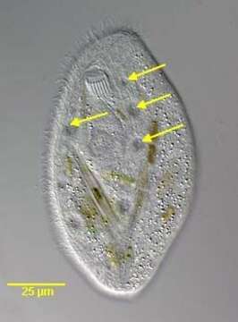
Ventral view of the chillodonellid ciliate, Pseudochilodonopsis polyvacuolata (Foissner and Didier, 1981). The cell is ovoid. The anterior end is drawn to the left as a bluntly pointed rostrum. The ventral surface is flat and the central dorsal surface is arched. There is a flattened narrow circumferential margin. Ciliature is restricted to the ventral surface except for a short dorsal brush. The 7 left somatic kineties are separated from 5 right somatic kineties by an unciliated postoral bare area. The lateral-most 5 left somatic kineties terminate at a right angle to short separate preoral kineties arranged in stair-step fashion from the cytostome to the tip of the rostrum. The medial two left somatic kineties are shorter. There are two short circumoral kineties. The cyrtos opens ventrally. The heteromerous macronucleus is approximately central with one adherent ovoid micronucleus. There are 7-10 contractile vacuoles each with a single ventral excretory pore (arrows). Collected from a freshwater stream with abundant pennate diatoms near Boise, Idaho;43° 34' 41.92" N 116° 08' 50.49" W. March 2006. DIC.
Included On The Following Pages:
- Life (creatures)
- Cellular (cellular organisms)
- Eukaryota (eukaryotes)
- SAR (Stramenopiles, Alveolates, Rhizaria)
- Alveolata (alveolates)
- Ciliophora (ciliates)
- Intramacronucleata
- Phyllopharyngea
- Phyllopharyngia
- Chlamydodontida
- Chilodonellidae
- Pseudochilodonopsis
- Pseudochilodonopsis polyvacuolata
This image is not featured in any collections.
Source Information
- license
- cc-by-nc
- author
- William Bourland
- provider
- micro*scope
- original
- original media file
- visit source
- partner site
- micro*scope
- ID