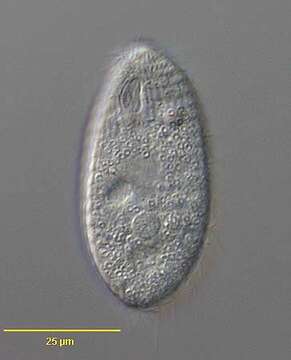
in vivo; ventral view
Description:
Portrait of the hymenostome ciliate, Dexiotricha granulosa (KENT,1881) FOISSNER, 1994, synonymous with Loxocephulus granulosus. The cell is ovoid, broadly rounded posteriorly and truncate anteriorly. Regular longitudinal kineties terminate at a subapical band of circumferential kineties demarcating a cilia-free truncate apical area or frontal plate. There is a single long caudal cilium. The oral aperture is small and difficult to visualize in vivo. It is located in the anterior quarter with an undulating membrane on the right (seen faintly here) and 3 membranelles (not seen here). The macronucleus is spheroid and located in the mid-cell. The contractile vacuole is seen here to the left of the macronucleus. The spherical micronucleus is not seen here. The cytoplasm contains many small refractile ring-shaped glycogen granules, which are diagnostic for the species (see detail images). Dexitricha is bactiverous. From freshwater pond near Boise, Idaho. Differential interference contrast.
Included On The Following Pages:
- Life (creatures)
- Cellular (cellular organisms)
- Eukaryota (eukaryotes)
- SAR (Stramenopiles, Alveolates, Rhizaria)
- Alveolata (alveolates)
- Ciliophora (ciliates)
- Intramacronucleata
- Oligohymenophorea
- Scuticociliatia
- Philasterida
- Loxocephalidae
- Dexiotricha
- Dexiotricha granulosa
This image is not featured in any collections.
Source Information
- license
- cc-by-nc
- author
- William Bourland
- provider
- micro*scope
- original
- original media file
- visit source
- partner site
- micro*scope
- ID