composite
Description:
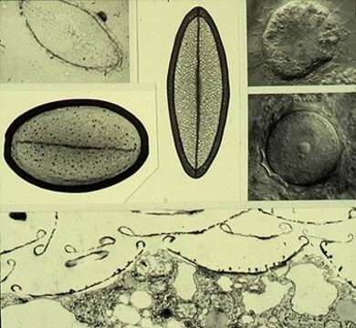
Composite image showing single scales (upper left). Upper left corner treated with hydrofluoric acid to show siliceous nature, vertical scale from trophic cell (Nomarski image upper right corner), larger scale from the cyst (middle right image). The lower image is a transmission electron micrograph of a thin section showing the scales adhering to the outer surface of the cell.
Included On The Following Pages:
- Life (creatures)
- Cellular (cellular organisms)
- Eukaryota (eukaryotes)
- Haptista
- Centroplasthelida (Centrohelid)
- Raphidiophryidae
- Raphidiophrys
- Raphidiophrys ambigua
This image is not featured in any collections.
Source Information
- license
- cc-by-nc
- author
- D. J. Patterson.
- provider
- micro*scope
- original
- original media file
- visit source
- partner site
- micro*scope
- ID